
01 October 2026
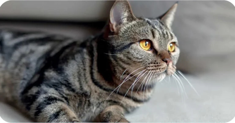
Carus presents the case of Barney, a 14-year-old cat with longstanding gastrointestinal signs.

Differentiating feline chronic inflammatory enteropathy from low-grade intestinal T-cell lymphoma (LGITL) can be challenging. Clinical signs, laboratory findings, ultrasonographic changes and even histopathological features can overlap.
Faecal calprotectin cannot distinguish between the two, but it can provide an objective measure of intestinal inflammation and help inform the next step. In Barney’s case, that proved particularly useful.
Barney, a 14-year-old neutered male European shorthair, presented with a four-week exacerbation of longstanding GI signs. His diarrhoea had become more frequent, he was vomiting daily and his appetite had reduced. He had also lost 400g over the previous five months.
Differential diagnoses included chronic enteropathy, intestinal lymphoma, pancreatitis and extra-gastrointestinal disease.
Routine blood testing identified increased ALT and ALP alongside marked hypocobalaminaemia, while PLI remained within normal limits. Abdominal ultrasonography revealed mild generalised thickening of the jejunum and ileum, relative muscularis layer thickening and prominent jejunal and colic lymph nodes, but no intestinal mass.
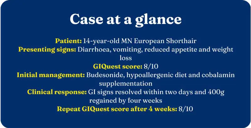
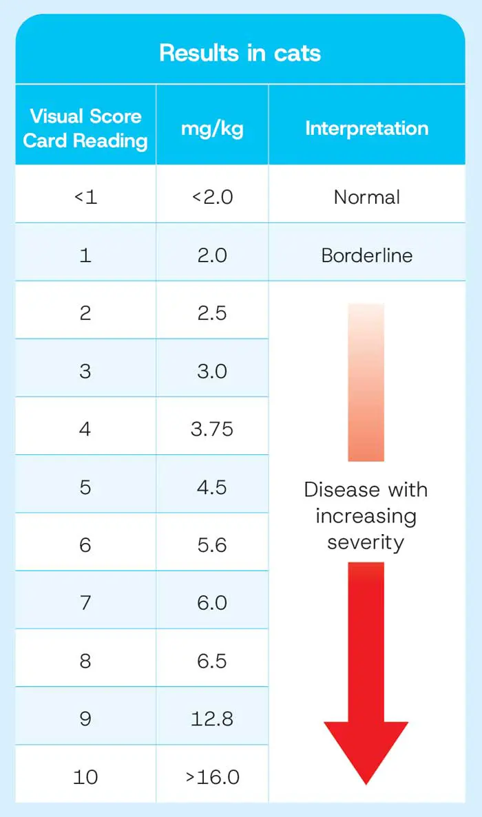
Barney’s initial GIQuest score was 8/10, supporting the presence of significant intestinal inflammation.
Although his vomiting, diarrhoea and inappetence resolved within two days and he regained 400g by four weeks, repeat testing showed his GIQuest score remained unchanged at 8/10.
Considered alongside the marked hypocobalaminaemia, muscularis thickening and lymphadenopathy, this persistent elevation supported further investigation despite the clinical improvement.
Endoscopy and intestinal biopsy were subsequently performed. Histopathology identified marked intraepithelial accumulation of monomorphic small lymphocytes, with emerging epitheliotropic lymphoma considered the most likely diagnosis.
A diagnosis of small-cell gastrointestinal T-cell lymphoma, also commonly termed LGITL, was reached and treatment with prednisolone and chlorambucil was started.
Following diagnosis, Barney was started on prednisolone at 5mg once daily alongside three cycles of chlorambucil at 4mg once daily for four days, repeated every 21 days. Following the second cycle, faecal calprotectin was reassessed using GIQuest.
This time, Barney’s GIQuest score had fallen from 8/10 to 4/10, reaching 1/10 two weeks later. The reduction provided an objective indication of treatment response and supported continuation of the planned protocol.
Definitive diagnosis of LGITL requires intestinal biopsy and histopathology, with additional tests such as immunohistochemistry or PCR for B- and T-cell clonality sometimes required.
In Barney’s case, repeat GIQuest testing identified persistent inflammation despite apparent clinical improvement, prompting progression to intestinal biopsy and definitive diagnosis. Subsequent reduction in his GIQuest score provided an objective measure of response once disease-specific treatment was underway.
GIQuest testing will now be repeated at future rechecks to assess gastrointestinal inflammation and potential relapse non-invasively.
GIQuest is a patient-side lateral flow test for faecal calprotectin, providing a non-invasive way to assess intestinal inflammation in dogs and cats.
Used alongside history, clinical examination and other diagnostic findings, serial testing can provide an additional objective measure when monitoring GI disease and help make the next step clearer when clinical signs alone may not tell the full story.
To explore more case studies and find out more about using faecal calprotectin in both feline and canine GI cases, visit GIQuest – Carus.
Alternatively, submit your own case study here.
* Case example based on a clinical case provided by Tracy Chavez Pino, BVetMed, MRCVS, PgCert Dent (WBIS) and Greg Dixon, MA, VetMB, CertWE,L PgC SADI, PgC SAM, PhD, MRCVS