1 Oct 2018
Rachel Hattersley provides guidance on assessing and managing these types of skin injuries.
Rachel Hattersley
Job Title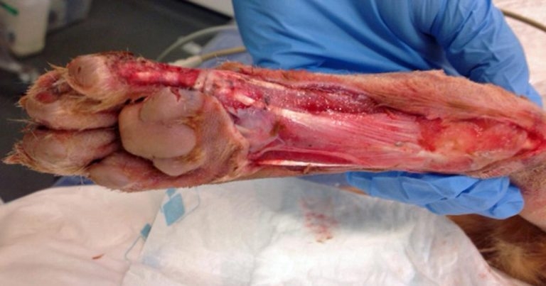
Figure 1. Degloving injury to the distal limb of a dog with exposure of underlying tendons and ligaments.
We all know that sinking feeling when the call comes in about a trauma patient just at the busiest moment in the day, and significant skin wounds are not uncommon in such patients.
Large skin wounds can require significant longer-term management that can impact patient welfare and also the owner, financially and emotionally. A degloving injury is a type of avulsion injury in which an extensive section of skin is completely torn off the underlying tissue, severing its blood supply (Figure 1).
This article will aim to provide the practitioner with a review of how to manage such injuries in a logical manner that optimises outcome.

The degree of trauma required to cause any severe skin injury will often – but not always – be significant.
While the temptation is to concentrate on the obvious external injuries, concurrent thoracic and abdominal trauma is not uncommon when avulsion injuries are present. In one study of 235 dogs experiencing severe blunt trauma, pulmonary contusions were reported in 58% of all patients and pneumothorax in 47% (Simpson et al, 2009).
The primary survey, performed on initial assessment of the patient, aims to assess the most essential body systems, such as respiratory, cardiovascular, urinary and neurological systems; prior to sedation or anaesthesia for other diagnostic investigations. Triage begins with initial owner contact to the practice. The reception team can aid in the prioritisation of patients by asking specific questions to owners during telephone contact to determine the severity of the situation.
On arrival at the practice, an initial assessment should be made to determine if the patient needs to be removed from its owner immediately for emergency treatment or if the patient can remain with the owner during the consultation. Once admitted, IV access should be obtained and the primary survey performed. Oxygen should be administered if evidence of respiratory compromise exists. Based on the results of the primary survey, IV fluid therapy can be given as required. The rate and type of fluids should be tailored to the individual.
Crystalloids are most commonly used as the first line fluid, but colloids should be considered if significant blood loss is suspected. It should be noted it is often more appropriate to give a bolus of fluids (5ml/kg to 10ml/kg) over a 15-minute period then reassess clinical parameters and give further boluses as required (based on changes in heart rate, respiratory rate, blood pressure and capillary refill time) rather than a set rate in ml/kg/hr for every patient.
Rapid analgesia is essential for the majority of patients presenting post-trauma. Analgesia should be tailored to the individual, but an opioid (and preferably a full mu-agonist, such as methadone) is usually indicated. Realistically, the detrimental effect of pain on normal respiration (such as fractured ribs) will usually far outweigh the respiratory depression caused by opioids. If an opioid alone is inadequate, other options for analgesia include either a ketamine constant rate infusion (CRI) or a lidocaine CRI.
IV paracetamol can also be considered in canine patients (10mg/kg as a slow IV infusion over 15 to 30 minutes). NSAIDs should be avoided in hypovolaemic, hypotensive patients as an increased risk of renal toxicity and gastric ulceration exists. These drugs can be introduced once the patient is stable and any volume deficits have been corrected.
Wound assessment usually requires deep sedation or anaesthesia, and, therefore, must be delayed until patient stabilisation is complete. Large wounds should be covered with a sterile dressing to prevent further contamination in the interim period. Once anaesthetised, the limbs should be carefully palpated for any evidence of fracture or instability.
If you have any suspicion of concurrent orthopaedic injury, orthogonal radiographic views should be obtained. Shearing injuries can often be associated with ligamentous damage to the tarsus/carpus and palpation of the joints should be performed. Stressed radiographs may be required for definitive diagnosis.
Wound assessment and initial management involves four steps:
| Table 1. Summaries of dressing options for degloving injuries | |||||
|---|---|---|---|---|---|
| Dressing type | What? | When? | Why? | Frequency of dressing changes | Points to note |
| Wet to dry dressing | Adherent. Sterile swabs soaked in saline and wrung out as much as possible. | During the inflammatory phase of wound healing in wounds that are highly exudative and contaminated with debris. This dressing should only be used during the initial debridement period as removal of the dressing can damage epithelial cells and capillary buds. | The exudate soaks through the swabs in to the secondary layer of the bandage. As the swabs dry out they adhere to the wound bed and when removed, debrides the wound bed. | Every 12 to 24 hours | These dressings are painful to remove and, thus, dressing changes require sedation or anaesthesia. |
| Polyurethane dressings | Non-adherent absorptive dressing that maintains a moist wound environment. | During both the inflammatory and proliferative phases of wound healing. | Improved patient comfort. The moist wound environment promotes autolytic wound debridement. | Depends on volume of exudate. | Ensure the dressing does not have an occlusive backing to prevent skin maceration. |
| Alginate dressings | Calcium alginate (derived from seaweed). | In highly exudative wounds. | Absorb exudate to create a hydrophilic gel at the wound surface, which traps bacteria and debris. | Need to be left in situ for approximately three days to allow the gel to form. | The gel will not form in a dry wound so avoid use in minimally exudative wounds. |
| Hydrocolloid dressings | Hydrophilic polymers. | On wounds producing only small volumes of exudate to promote epithelialisation. | As the gel absorbs exudate, it liquefies to form a viscous gel at the wound surface, which maintains a moist wound environment and promotes autolytic debridement. | Dependent on the degree of exudate. | |
| Silver dressings | Either as elemental silver, an inorganic compound or an organic compound. Silver needs to become a positively charged ion to be effective as a bactericidal agent. | In infected wounds. | Silver ions are highly reactive and affect multiple sites within bacterial cells, ultimately causing bacterial cell death. They bind to bacterial cell membranes, causing disruption of the bacterial cell wall and cell leakage. Silver ions transported into the cell disrupt cell function by binding to proteins and interfering with energy production, enzyme function and cell replication. | Dependent on the degree of exudate. | |
A detailed description of the management of specific orthopaedic injuries is beyond the scope of this article. However, as discussed previously, concurrent ligamentous injuries are common in companion animals that have sustained a degloving injury. As the majority of such injuries are “open”, the use of synthetic materials to reconstruct prosthetic ligaments or augment autogenous tissues can be challenging due to the high risk of implant-associated infection.
Transarticular external skeletal fixators are often used to maintain the affected joint at a functional angle while permitting fibrosis of the damaged soft tissue structures. Should this not be sufficient to stabilise the joint, pancarpal or pantarsal arthrodesis can be considered once the skin wounds have healed. Immobilisation of the affected joint is also often beneficial for wound healing if free skin grafting is used.
The location and configuration of the wound will determine whether it is necessary to attempt exploration of the wound and immediate wound closure.
Penetrating or shearing wounds of the thorax and abdomen should be explored and closed over drains (ideally closed suction drains), after appropriate and thorough lavage (Figure 2). Any obviously dead skin should be removed prior to closure and the owner should be made aware further skin loss may occur during the healing process, which may require further surgical debridement.
As the majority of degloving or shearing injuries occur on the appendicular portion of the skeleton, it is often preferable to have a period of open wound management before attempting either delayed primary wound closure (if adequate skin is available) or use of a technique to mobilise skin to the area. This will allow time for sequential debridement of the wound, thus any areas of skin of questionable viability can be left in situ and debrided, if required.
It is important to remember, especially at early points during wound management, it is likely dressing changes will be required on a daily basis. Ensure this is done in an aseptic manner. As daily general anaesthesia or sedation is likely to be required, it is vital patient nutrition be considered to ensure an adequate calorie intake is maintained. Analgesia should also be tailored to the requirements of the patient.
The subject of antibiotic therapy remains somewhat controversial. Inevitably instincts often lead to the administration of broad spectrum antibiotics in such cases. However, once a healthy granulation bed has formed, this is unnecessary. It is usual for there to be exudate from open wounds and this should not immediately mean antibiotic therapy; such actions will only select for a more resistant resident population of bacteria on the surface of the wound.
Red flags include an increase in the amount of discharge or change in the nature of the discharge. In such cases, a swab should be taken in an aseptic manner and submitted for bacterial culture. Use of topic therapy, such as medical grade Manuka honey or silver sulphasalazine cream, should be considered before systemic treatment.
With appropriate wound management, clinicians will find the majority of wounds will heal by second intention without further intervention. However, as wound contracture on the distal limb will be limited, this can take some considerable time as wound healing is reliant on epithelisation alone.
Depending on the location on the limb, closure of larger wounds can either be achieved by axial pattern flaps (if an appropriate option exists) or by free skin grafting (most commonly used on the distal limb).