
29 Sept 2026
Kieran Borgeat BSc, BVSc, MVetMed, CertVC, FHEA, DipACVIM, DipECVIM-CA (Cardiology), FRCVS explores the various stages of this issue and how clinicians can use diagnostics to intervene before signs of heart failure appear.

Kieran Borgeat
Job Title
Image: stokkete / Adobe Stock
Mitral valve disease (MVD) is an important welfare problem for dogs, affecting up to 3.5% of all dogs in the UK (Mattin et al, 2015), but with a significantly higher prevalence in smaller breeds (less than 15kg) older than six years of age.
The typical early warning sign of MVD is an audible murmur, detectable at routine veterinary health checks. Many dogs remain apparently healthy for years, because of a long preclinical disease course; however, early signs in this “asymptomatic” phase may be subtle and attributed to the ageing process by many owners.
In addition, many dogs are not exercised to their capacity, so early evidence of cardiovascular impairment is missed. Interestingly, in the 2016 EPIC study – which will be discussed further on in this article – dogs that were reported by owners to be asymptomatic had an improvement in quality of life score on treatment: what had the owners missed?
In time, the worst affected dogs develop clinical signs of reduced cardiac output (exercise intolerance and prolonged recovery) and congestive signs (pulmonary oedema). Coughing is common, but is more a sign of increased atrial pressure and triggering of cough mechanoreceptors in the principal bronchi than oedema. Some dogs present with syncope, often associated with a hyperdynamic left ventricle (severe mitral regurgitation) and inappropriate activation of a cardio-depressor reflex, leading to bradycardia and hypotension.
In later disease, dogs can develop complicating factors such as pulmonary hypertension or arrhythmias such as atrial fibrillation. Sudden death is reported to occur in up to 50% of cases, associated with full-thickness atrial tears or complex arrhythmias.
However, more than half of dogs die with the disease rather than because of it. Identifying those more progressive cases is a crucial part of clinical practice, to prioritise where to focus diagnostics and treatment to best help improve welfare.
We understand that many dogs with MVD have a relatively benign disease course. As is common in 21st century practice, MVD is considered on a spectrum of disease stages, using the ABCD model (Figure 1).
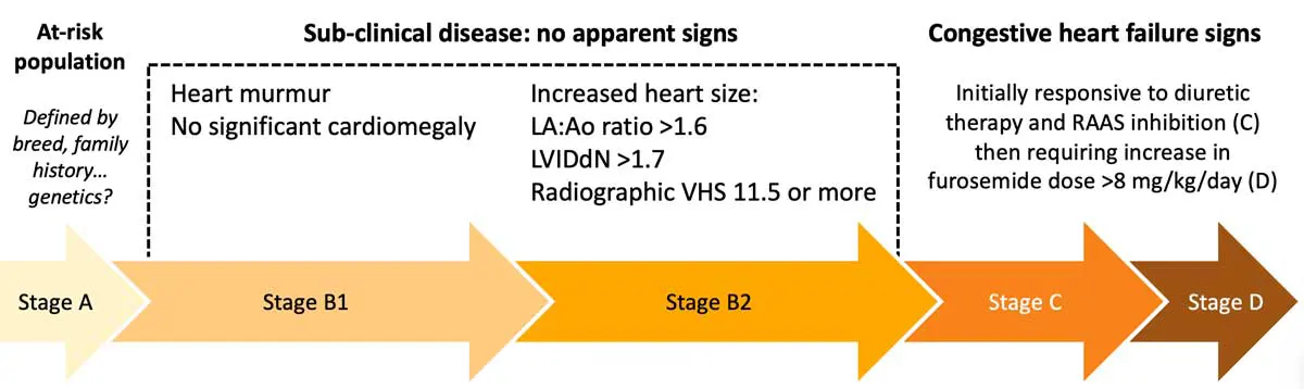
Stage A is difficult to accurately define. In humans, similar risk profiles are established using details from family history and genetic profiling.
In veterinary practice, we often use breed as a marker. The cavalier King Charles spaniel, Chihuahua, cocker spaniel, poodle, bichon frisé, terrier breeds, miniature dachshund and their cross-bred forms (cavapoo, cavachon and numerous others) are considered to be at risk, but often we lack a detailed family history. Although steps towards better understanding the genetics of MVD in dogs (particularly in the cavalier King Charles spaniel) are being taken, we still do not know enough to begin considering the reliable use of genetic tests in decision making.
Due to this, stage A is more a philosophical stage than a practical assessment of MVD risk in dogs.
Stages C and D are overtly affected by clinical signs. In stage C, dogs require loop diuretic therapy (furosemide, torasemide) to control clinical signs of tachypnoea caused by pulmonary oedema. In time, the nephron adapts and dogs become reliant on increasing doses of medical therapy.
Once they are on standard treatment (pimobendan, spironolactone, an ACE-inhibitor) plus a loop diuretic dose equivalent to furosemide at 8mg/kg/day or more, they are considered refractory: stage D. Here, complicating factors such as pulmonary hypertension and arrhythmias are more common. Over the past 20 years, numerous large-scale studies have shown a survival time of 9 to 12 months for dogs once diagnosed with stage C disease. Identification of stage C relies on using history, physical examination and imaging findings to confirm the diagnosis of MVD and congestive signs.
Stage B is the more challenging phase to identify. In addition, stratifying dogs into stage B1 or B2 is a key part of management of MVD. Since 2016, we have considered that treatment with pimobendan (0.25mg/kg to 0.3mg/kg orally every 12 hours) in stage B2 represents standard of care. In the EPIC study (Boswood et al, 2016), dogs in the subclinical stage that had cardiomegaly on both echocardiographic and radiographic measures were randomised to receive either pimobendan or a placebo (identical in packaging and flavour, but not containing the active drug). Data showed several important improvements in the pimobendan-treated group, when compared to the placebo group:
Large disease registry data (The LOOK-Mitral Registry; Borgarelli, 2024, personal communication) show that approximately 70 per cent of dogs in stage B2 will never develop overt clinical signs of heart failure.
Treating every older, small-breed dog that has a heart murmur detected is, therefore, only going to help about one in three dogs, but puts a large financial burden on owners.
Arising from this, we need to consider how we identify the dogs that will benefit from treatment in this subclinical phase, so that our time and owner resources are best used.
Easy, right? We just need to perform echocardiography on all older, small-breed dogs with a heart murmur to accurately measure left atrium (LA) to aortic root (Ao) ratio and normalise left ventricular internal diastolic diameter (LVIDd) to bodyweight.
Well, for cardiology specialists or advanced practitioners, we might consider this sort of echo to be easy. The reality is that alignment of a 2D image plane within a complex 3D structure and positioning the measurement cursor poses a risk of inconsistency – particularly with the LA:Ao ratio.
This metric of heart size, regularly identified as a key decision maker, has a wide variation in repeatability even between specialists and is often over-measured by less experienced clinicians. Compound error from under-measuring aortic size above the sinus of Valsalva, combined with over-measurement of atrial diameter by including the orifice of a pulmonary vein, means that misclassification is easy and over-treatment can result.
Perhaps, more practically important – that would be an awful lot of dogs to scan.
So, a simple, accessible method to narrow down the number of dogs in which to perform an echo would be beneficial. The simplest method to stratify risk of a dog with a heart murmur having stage B2 MVD is heart murmur intensity.
The system of murmur grading on a I to VI scale of intensity is traditionally used, but can be practically interpreted as soft, moderate or loud/thrilling (Table 1). A moderate intensity murmur should trigger imaging of the heart, not prompt the prescription of pimobendan.
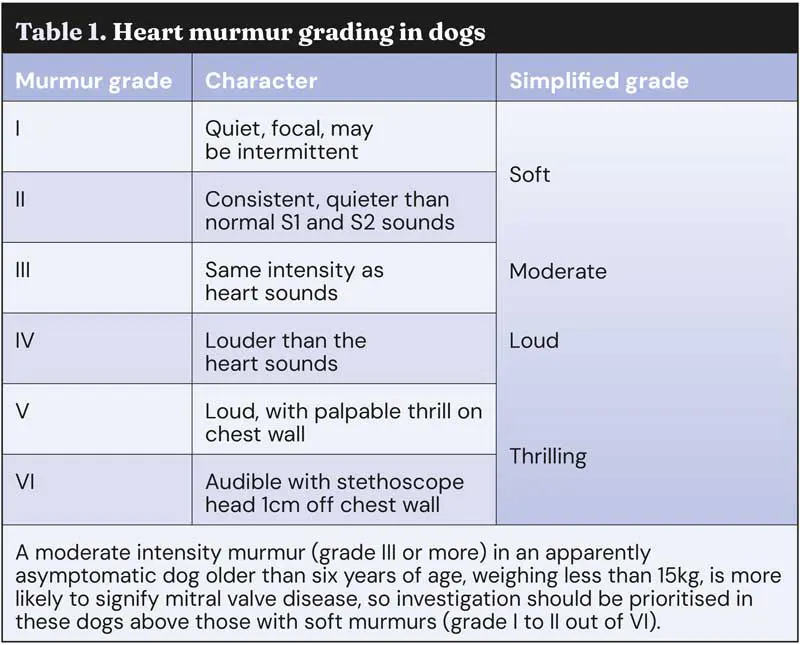
Grading heart murmurs consistently is more of a challenge than many anticipate. Variation in murmur grade between vets can be high, and learning auscultation and murmur assessment is difficult: everybody experiences sound in a different way, and people’s hearing changes with age, not to mention the difference in sound quality between different stethoscopes.
Of course, conformation of the patient, compliance and even haircoat thickness have a role to play in making auscultation of heart murmurs more complex. Over time, the changing landscape of clinical vet student teaching in the UK means that almost all universities now have cardiology as a non-core rotation, if it features at all in rotations. This relies on more peer-to-peer teaching of auscultation in the primary care setting, putting more pressure on general practitioners to have a confident and reliable approach to murmur grading.
The use of electronic stethoscopes can help by providing a concurrent phonocardiogram – a visual representation of heart sounds. In combination with a recently released smartphone app (EkoVet+), which uses an artificial intelligence (AI) system to grade canine mitral murmurs as reliably and repeatably as an international panel of five cardiology diplomates (Figure 2), clinicians can reach for additional input on their findings. Self-testing by comparing the murmur grade assessed by the clinician auscultating the patient with the output from the AI system is an opportunity for training in heart murmur grading that is accessible in the consultation room.
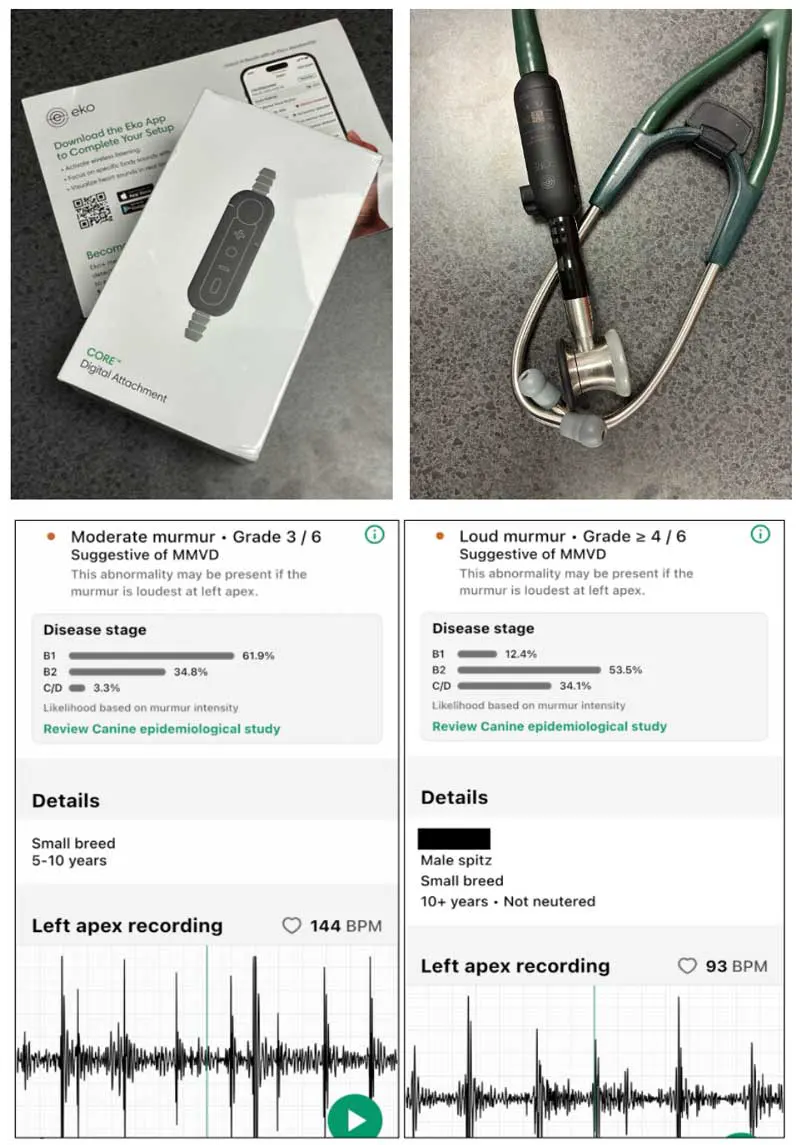
We can refine our case selection for pimobendan treatment beyond the grading of heart murmurs. Some dogs with a loud murmur have mild disease. Even with the utility of reliable auscultation, this is only the first step to select dogs that will be most likely to benefit from drug intervention.
Another accessible test outside of referral clinics is the measurement of NT-proBNP, a cardiac biomarker indicating cardiac stretch or stress. Various commercial reference laboratory assays are available for quantitative measurement in dogs, and in-house assays have recently become available (more stringent sample handling is often necessary; if in doubt, run within an hour of sampling).
Alongside this, most vets have easy access to radiography, allowing us to assess heart size on thoracic radiographs.
In dogs at risk of MVD, with a moderate or loud intensity heart murmur on the left side, imaging of the heart as a next step is probably the most efficient route to identifying stage B2.
If echocardiography is not an option because of financial cost, or limited access to a person locally with sufficient experience to provide reliable measurements, then a right lateral thoracic radiograph should be obtained.
Vertebral heart sum (VHS), although prone to wide inter-individual variation of numerical measurement, as well as name (the S is often defined as scale or size), is reported to be the most sensitive method to detect radiographic stage B2 MVD. Method is important, and measuring VHS should be performed as carefully as any echo parameter (Figure 3). If VHS measures 11.5 or higher, stage B2 MVD is likely, and pimobendan can be confidently prescribed.
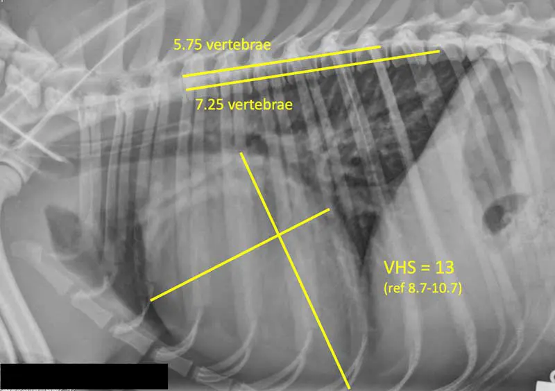
Beware the use of alpha-2 agonist drugs as part of sedation plans for obtaining radiographs: even at low doses, they will increase heart size significantly because of their effect on loading conditions, so over-diagnosis will be frequent if dexmedetomidine or other drugs in this class are administered.
In addition, make sure the radiographic view is not over-rotated, as this will reduce the reliability of VHS measurement.
Where VHS is equivocal (between 10.5 and 11.5), NT-proBNP may be a good test to support the decision to prescribe pimobendan.
Various studies have been performed to provide cut-offs for NT-proBNP that discriminate between subclinical MVD stages, but different assay types, breed differences in normal NT-proBNP concentration in circulation, and up to 50% day-to-day variability for individual dogs all contribute to wide “grey zones” in the guidance.
Generally speaking, a dog with equivocal radiographic heart size and an NT-proBNP of more than 1,000pmol/L is likely to have stage B2 MVD. However, data from cavalier King Charles spaniels using a protocol of moderate intensity murmur, VHS higher than 11.5 and NT-proBNP of more than 1,000pmol/L still misclassified one in four dogs (Wesselkowski et al, 2023).
Where an echocardiogram performed by an experienced clinician is available, it should always be considered the better test.
A subset of dogs with apparently asymptomatic MVD will have an improved quality of life and longevity before the onset of heart failure signs if we can identify those with cardiomegaly and prescribe pimobendan.
Diagnostics, starting with a good auscultation and considered grading of the heart murmur, but also employing the use of cardiac imaging, allows us to avoid over-treatment of the 70% of dogs with early MVD that never progress to develop clinical signs.
In the absence of echocardiography performed by a suitably experienced operator, thoracic radiographs combined with NT-proBNP probably represent the best set of tests to identify dogs where treatment should be prioritised.
Kieran Borgeat qualified from the University of Bristol and worked in primary care practice before undertaking a cardiology residency. He became a diplomate in 2014 and has worked in private referral and academic practice since then. Kieran holds the American and European board certification, and is an RCVS-recognised specialist in veterinary cardiology. He is head of the cardiology service at Bristol Vet Specialists, where he has developed one of Europe’s leading hybrid and interventional cardiac surgery units, and is producer and co-host of The Animal Heartbeat podcast.