26 Aug 2021
Guillaume Albertini and Fabio Stabile update readers on information published surrounding the most common of these conditions that have an impact the central nervous system in canine patients.
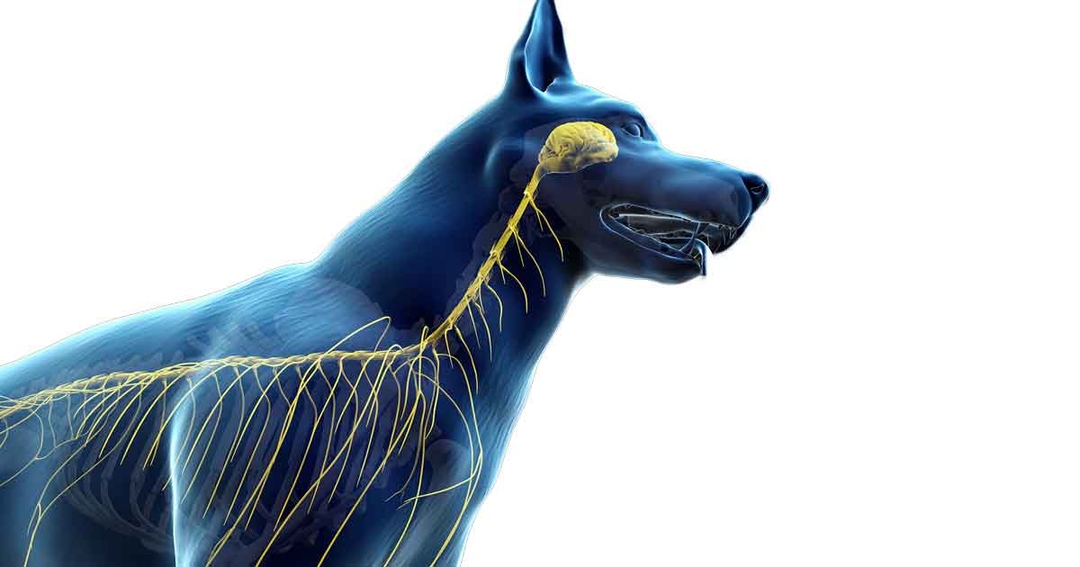
Image: © SciePro / Adobe Stock

Over the course of the past 50 years, neurological disorders affecting the CNS of small, large and exotic animals have been gradually documented. New conditions or additional information regarding known diseases are brought to the veterinary literature on a daily basis.
The purpose of this review is to update veterinary surgeons on information published over the past five years on the most common neurological disorders affecting the CNS.
Canine cognitive dysfunction (CCD) has been considered the canine analogue of human Alzheimer’s disease1-3. CCD is common in older dogs, particularly those older than eight years1-3. Estimated prevalence of CCD generally varies between 14% and 35% of the domestic canine population depending on publications1-5. However, many of the reported prevalence likely underestimates how common this disorder is.
The pathophysiology of CCD is multifactorial and complex. Progressive accumulation of neurotoxic protein Aβ in neurons, microglia and astrocytes is a constant feature of CCD and Alzheimer’s disease. In addition to Aβ deposition, vascular disease, numerous cellular (microglia and astrocytes) and biochemical aberrations (neuronal mitochondrial dysfunction, glutamate-mediated excitoxicity) contribute to progressive cognitive decline6-10.
The disorder is characterised by four main features reported by owners:
Dogs with CCD typically show evidence of forebrain dysfunction on clinical examination1-3,10-12. These patients show abnormal mentation and often respond inappropriately to their environment (dementia)1-3,10-12. Many dogs with CCD circle constantly in the examination room, and either do not respond or respond inappropriately to visual and auditory stimuli1-3,10-13. Studies found a significant association between idiopathic epilepsy and early-age onset of CCD in dogs14-16.
The diagnosis of CCD is based on signalment, history, and clinical features1-3. Because patients with suspected CCD are generally elderly, and the clinical features of CCD are numerous and often non-specific, it is important to rule out geriatric disorders that may mimic signs of cognitive impairment1-3,17-21.
Laboratory findings (comprehensive biochemistry, haematology) are usually normal in patients with CCD (unless a concurrent age-related condition is identified). In particular, a differential diagnosis of a forebrain tumour is typically a top consideration in an elderly dog with behavioural changes. Organ dysfunction (such as liver, kidneys) and discomfort may also lead to clinical signs that may be confused with cognitive impairment1-3.
The only imaging modality of practical use is MRI. MRI features reported in CCD are cerebrovascular diseases (ischemic lesions, microhaemorrhages and occasionally macrohaemorrhages); cerebral atrophy (suspected when the interthalamic adhesion thickness is smaller than 5mm); meningeal thickening; ventricular dilatation; and gliosis18-20.
Contrary to the human Alzheimer’s disease, CCD is usually less severe1-3. Dogs with CCD usually have a good quality of life, though cognitive function may continue to decline1-3,21. No consensus exists regarding treatment of CCD. The main purpose of the treatment is to slow the disease progression. Treatment may rely on three main pillars: dietary modulations, a variety of nutraceutical supplements and cognitive enrichment22-28.
Dietary modulation in dogs includes an increased ratio of medium-chained triglycerides (MCTs)23-25. MCTs are recommended in dogs with CCD, either as part of a formulated diet or as a supplementation to a commercial well-balanced diet23,24. The CCD brain has an impaired ability to use glucose, the brain’s main energy source. MCTs provide an alternative energy source for the brain in cognitively impaired patients26.
Nutraceutical supplements and drugs may be used to regulate some clinical signs of CCD (such as melatonin and dog-appeasing pheromones to attempt regulation of sleep-wake syndrome and anxiety)28. No drug is showed to improve cognitive function in these patients.
Cognitive enrichment – such as regular exercise, social interactions and introduction of new toys – has been shown to improve cognitive function in dogs with CCD and prevent delay of cognitive decline in dogs as they age22-28.
Chiari-like malformation (CM) is a complex developmental condition of the skull and craniocervical vertebrae, and is characterised by a conformational change and overcrowding of the brain and cervical spinal cord, particularly at the craniospinal junction29,30. Obstruction to CSF flow can result in formation of fluid-filled cavitation in the spinal cord, called syringomyelia (SM), and secondary neuropathic pain31.
The rising availability of MRI has allowed CM and SM to be more commonly diagnosed and understood in veterinary medicine32,33. Published studies investigating CM and syringohydromyelia in the cavalier King Charles spaniel (CKCS) breed suggested that by five years of age, 70% of the population have MRI evidence of SM33,34.
CM and SM still represent a challenging diagnosis as a degree of CM and SM is “expected” in predisposed breeds such as the CKCS, but may not warrant treatment34. The clinician should aim to answer the question when and how should CM and SM be treated? Treatment should be initiated when the clinical signs impact quality of life35.
Clinical signs often associated with CM and SM are spontaneous vocalisation, spinal discomfort, head and ear rubbing or scratching, phantom scratching (that could mimic ear scratching), aversion to touch and behavioural changes (such as becoming anxious, timid, aggressive or withdrawn)30,31,36,37. These clinical signs are not specific of CM and SM. Therefore, even in predisposed breeds, comprehensive investigations including MRI and, if safe to perform, CSF collection and analysis are recommended to rule out other conditions that could present similar clinical signs (such as inflammatory diseases of the CNS and intervertebral disc diseases)38.
Clinical signs will progress in approximately 75% of dogs33,35. Despite progressive signs, many dogs with discomfort and phantom scratching respond to medical treatment, and are considered to have an acceptable quality of life33,35.
Medical treatment relies mostly on oral analgesic medications and other, currently unlicensed, medications. These are summarised in Table 1 33,35,39-44. Surgical management is more clearly indicated and more likely to be successful in dogs with CM (with or without SM) that have not responded or responded incompletely to medical treatment33,35.
| Table 1. Common medications used for management of Chiari-like malformation and syringohydromyelia pain-related syndrome33,35,39-44 | ||
|---|---|---|
| Molecule | Dose | Adverse effects |
| NSAIDs | As per manufacturer recommendations | Gastrointestinal signs (vomiting, diarrhoea). Not recommended for patients with kidney disease. Contraindicated in patients receiving corticosteroid anti-inflammatories |
| Paracetamol | 10mg/kg/8 to 24 hours | Risk of renal, hepatic failures, gastrointestinal signs, haematologic toxicity (methaemoglobinaemia), keratoconjunctivitis sicca |
| Gabapentin | 10mg/kg/8 hours to 20mg/kg/8 hours | Sedation/ataxia, gastrointestinal signs and increased appetite described |
| Pregabalin | 5mg/kg/12 to 24 hours to 10mg/kg/12 to 24 hours | Sedation and ataxia at first initiation of the treatment |
| Amantadine | 3mg/kg/24 hours to 5mg/kg/24 hours | Sedation and ataxia. Avoid in patient with glaucoma, hepatic disease, renal disease, congestive heart failure, atopic dermatitis or seizures |
| Omeprazole | 0.5mg/kg/12 to 24 hours to 1.5mg/kg/12 to 24 hours | Reported side effects include nausea, diarrhoea, constipation and skin rashes |
| Prednisolone (corticosteroids anti-inflammatories) | 0.5mg/kg/24 hours then decreased to lowest possible dose | Polyuria, polydipsia, polyphagia leading to weight gain, vomiting, diarrhoea, muscle loss, panting, restlessness, poor hair coat quality |
Craniocervical decompression, ventriculo-peritoneal shunting and syringo-pleural or subarachnoid shunting are three reported surgical options available. However, none are entirely satisfactory and may present severe comorbidity risks45,46. Acupuncture has also been described as a useful adjunctive therapy in cases where clinical signs are poorly controlled or that present severe side effects of treatment.
A series of consensus statement articles published in 2015 by the International Veterinary Epilepsy Task Force (IVETF) provided precise guidelines on the definition, diagnosis, treatment and prognosis of canine idiopathic epilepsy. The IVETF recommendations are still the most updated consensus on the management of idiopathic epilepsy47-51.
Recent publications have therefore aimed to evaluate quality of life of patients suffering from idiopathic epilepsy, evaluate the owner’s perception of his or her pet and research new potential adjunctive treatments (such as dietary changes or herbal treatment) to the conventional anti-epileptic medication51-54.
Quality of life – as defined by the World Health Organization – describes a state of complete physical, mental and social well-being, and not merely the absence of disease and infirmity54. Quality of life is always difficult to completely appreciate in canine patients. Idiopathic epilepsy as a chronic disease puts a heavy burden on quality of life, whether because the patient is still suffering from epileptic seizures or is suffering from quality of life-limiting side effects51.
With idiopathic epilepsy being a multifactorial brain disease, new treatment strategies should reflect this in a more multimodal approach to idiopathic epilepsy management.
A recently developed diet based on MCTs has shown encouraging results as an adjunctive treatment to conventional antiepileptic medication to control epileptic seizure frequency and severity, in a small cohort of 21 patients diagnosed with idiopathic epilepsy55. During the six-month study, 3 patients (14%) were epileptic seizure-free, 7 patients (33.3%) had a reduction of seizure frequency greater than 50% and 5 patients (23.8%) had a reduction of seizure frequency less than 50%.
It is important to note that out of the 21 patients included, 6 (28.5%) did not appear to show any response to the treatment. These results must therefore be interpreted with caution due to the small sample study regarding the efficacy of this diet treatment. In addition, MCTs supplementation could also be effective to improve cognitive abilities in canine epilepsy56.
Previous research has focused on clinical aspects of epileptic seizure management on dogs with idiopathic epilepsy, with little attention given to the emotional and logistical challenges for their owners57,58. The commitment required to care for a dog with idiopathic epilepsy and the lifestyle changes made by their owners may be greater than estimated.
Medication compliance is one of the key features for epilepsy management. A recent study assessing owners’ compliance in canine idiopathic epilepsy showed that only a fifth of the studied population was 100% compliant58. In the same study, an idiopathic epileptic dog would miss a median of six days of treatment per cycle of treatment58. Also, patients receiving polytherapy (greater than one antiepileptic medication) showed a higher compliance rate than patients on monotherapy58.
These studies highlight the key role of veterinary surgeons in guiding and supporting the owners of epileptic patients to ensure a correct and consistent administration and monitoring of antiepileptic treatment.
A wide variety of inflammatory conditions can affect the CNS of dogs. For some conditions, an infectious agent (bacteria, fungus, virus, protozoal agent) can be identified59-61.
In the UK, the two most common agents responsible for infectious-mediated meningoencephalomyelitis in dogs are Toxoplasma gondii and Neospora caninum (protozoal agents)61. In the UK, in dogs diagnosed with a confirmed meningoencephalomyelitis, the prevalence of confirmed infection (based on PCR) by T gondii and N caninum is reported at 0.25% and 2.25%, respectively61.
When no infectious agent can be identified, the condition is suspected to be an immune-mediated inflammatory disease of the CNS and often falls under the umbrella term meningoencephalomyelitis of unknown origin, as a definitive diagnosis cannot be reached unless histological analysis is performed59,60,62,63.
The typical clinical features of the disease are an acute or chronic onset (sometimes insidious) of multifocal neurological signs (forebrain, brainstem, cerebellum, spinal cord) in young adult to middle-aged patients. The diagnosis relies on comprehensive blood work, MRI, comprehensive CSF analysis (including nucleated cell count, cytology and protein concentration) and exclusion of infectious agents exposure via antibodies serology titres, PCR on CSF and/or blood or CSF culture and sensitivity, whether indicated59,60,62,63.
Treatment and prognosis are currently the main field of research for inflammatory diseases of the CNS. Long-term immunosuppressive doses of corticosteroids slowly tapered over the course of several months are the first line treatment64-67. Due to the variable and sometimes poor response to corticosteroids monotherapy, as well as frequent severe and unacceptable side effects, a number of immunosuppressive medications have been evaluated as adjunctive treatments68-73. Table 2 summarises the different treatment protocols elaborated and the associated reported median survival times59-72.
| Table 2. Published treatment protocols for dogs with meningoencephalomyelitis of unknown origin and associated survival time59-72 | |||
|---|---|---|---|
| Molecule | Dose | Side effects | Median survival time |
| Prednisolone monotherapy | 0.5mg/kg/24 hours to 3mg/kg/24 hours | Polyphagia, polyuria, polydipsia, muscle atrophy, hepatopathy, skin disease (calcinosis cutis, poor hair coat quality) | 28 to 602 days |
| Prednisolone and cytarabine (constant rate infusion [CRI] plus SC injections) | Prednisolone: 0.5mg/kg/24 hours to 3 mg/kg/24 hours Cytarabine CRI: 100mg/m2 to 300mg/m2 over 8 to 24 hours then SC injections of 50mg/m2/12 hours over 48 hours repeated every 3 weeks for 3 cycles then extended to every 4 weeks for 3 cycles then further extended for 3 cycles |
Prednisolone-related side effect plus myelosuppression, gastrointestinal signs, transient post-treatment lethargy, dysphagia, limb tremors | 26 to 1,063 days |
| Prednisolone and ciclosporin A | Prednisolone: 0.5mg/kg/24 hours to 2mg/kg/24 hours Ciclosporin A: 3mg/kg/12 to 24 hours to 15mg/kg/12 to 24 hours |
Prednisolone-related side effect plus hypertrichosis, transient lymphopenia, vomiting, life-threatening anaemia | 236 to 1,345 days |
| Prednisolone and mycophenolate mofetil | Prednisolone: 0.5mg/kg/24 hours to 2mg/kg/24 hours Mycophenolate mofetil: 10mg/kg/12 hours to 20mg/kg/12 hours |
Prednisolone-related side effect plus gastrointestinal signs, sporadic infections, pancreatitis | 250 to 731 days |
Currently, oral prednisolone associated with cytosine arabinoside (cytarabine) administration provide the longest median survival time66,72. However, cytarabine administration protocols are often reported as time-consuming and financially challenging by some owners.
A recently published study compared two different prednisolone and cytarabine protocols73. This prospective study compared survival time, recurrence rate, and side effects of the standard cytarabine and prednisolone administration (as summarised in Table 2) in 42 dogs, with the administration of cytarabine constant rate infusion at the moment of diagnosis, followed by prednisolone treatment alone in 41 dogs73.
The results revealed that no difference existed regarding recurrence rate between the two protocols73. However, survival times could not be compared as most of the patients were alive at the end of the inclusion period for this study73.
In the UK, steroid-responsive meningitis-arteritis (SRMA) is the most common non-infectious inflammatory condition encountered in young dogs74. Diagnosis is often reached based on typical history (lethargy, cervical discomfort); signalment (young beagle, Bernese mountain dog, boxer); absence of neurological deficits; evidence of inflammatory condition of the CNS and exclusion of infectious CNS diseases74,75.
Although a specific trigger for SRMA is rarely identified, extensive workup (including synovial fluid examination) should be performed76. A correlation between SRMA and immune-mediated polyarthritis has been found76.
Conventional treatment for SRMA usually relies on immunosuppressive doses of corticosteroids74,75. Treatment with immunosuppressive doses of prednisolone (2mg/kg/24 hours to 4mg/kg/24 hours) usually results in rapid improvement. SRMA patients should then be slowly weaned off prednisolone over three to six months74,75.
Current research aims to identify adjuvant therapies to the corticosteroid treatment and aims to treat relapses of SRMA77,78. A recent publication described the use of azathioprine as a safe and efficacious adjuvant to corticosteroid treatment77. Cytarabine infusions have also been successfully described to treat recurrence of SRMA78.
Severe head trauma is associated with a high degree of morbidity and mortality in humans and animals. Brain injury in dogs is most often due to road traffic collisions; other causes include animal bites, falls and missile injuries (such as gunshot wounds)79.
Traumatic brain injury (TBI) has been documented to occur in 25% of severe blunt trauma cases in dogs79. TBI refers to injury to the CNS structures. TBI can be divided into primary brain injuries that occur immediately following the head trauma (such as intracranial haemorrhage and oedema) and that initiate a cascade of biochemical processes that result in secondary brain injuries79,80.
Direct brain parenchymal damage associated with primary brain injury is generally beyond the control of the veterinary surgeon in charge of the case. Currently no consensus exists on the treatment of severe TBI. However, it is of utmost importance to prevent increase in intracranial pressure (ICP), prevent damage to vital brainstem structures and provide adequate analgesic treatment79-81.
Initial physical assessment of the severely brain-injured patient should focus on imminent life-threatening abnormalities79. Many patients suffering from head trauma present in a state of shock (such as hypovolaemic shock). These patients should be stabilised with adequate IV fluid therapy before any neurological assessment.
A hypovolaemic patient with minimal brain injury will display signs of obtundation, mimicking a severe brain injury. Oxygen therapy is recommended for most acute brain-injured patients79-82.
Once the patients are stabilised from the initial shock, they should be assessed for injuries to the CNS, but also to other body systems (such as the respiratory, urinary and musculoskeletal systems).
MRI and CT are appropriate imaging modalities for TBI83-88. CT is often the preferred modality for imaging over MRI as its imaging acquisition speed is faster. Both MRI and CT images can provide prognostic indicators83-88. For example, the recently developed Koret grading system provides an objective scoring system based on CT imaging (Table 3)85,86.
| Table 3. CT findings constituting the Koret CT score and their scores86 | |
|---|---|
| Parameters | Score (points) |
| Haemorrhage | 1 |
| Midline shift or lateral ventricle asymmetry | 1 |
| Cranial vault fracture | 1 |
| Any caudo-tentorial lesion (hypodensity, haemorrhage, fracture) | 3 |
| Depressed fracture | 1 |
| Sum score | 0-7 |
| Caudo-tentorial lesions refer to lesions in the area caudal to the tentorium cerebelli, containing the cerebellum, cerebellar peduncle and medulla oblongata. | |
The Koret grading system is a point system (out of 7) assessing the following CT imaging features: haemorrhage, midline shift or lateral ventricle asymmetry, cranial vault fracture, caudo-tentorial lesions (hypodensity, haemorrhage, fracture) and depressed fracture86. The score is significantly associated with short-term and long-term survival of TBI dogs, and a cut-off of 3 points predicted survival with 85% sensitivity and 100% specificity86.
Medical treatment should be targeted to prevent elevation and/or decrease ICP79,81,82. Several physical therapies and medications have been described, and are summarised in Table 4.
| Table 4. Emergency management of increased intracranial pressure79,81,82 | ||
|---|---|---|
| Purpose | Procedure/medication | Recommendations |
| Minimise increases in intracranial pressure | • Elevate head at 30° angle • No jugular sampling or compression |
|
| Maintain cerebral blood perfusion | • Appropriate IV fluid therapy • Oxygenation (flow-by, nasal oxygen catheter, transtracheal catheter…) |
Assess patient for signs of hyperhydration (crepitus, tachypnoea…) |
| Decrease oedema | Corticosteroids: dexamethasone 0.15mg/kg/12 to 24 hours to 0.24mg/kg/12 to 24 hours IV followed by prednisolone 1mg/kg/24 hours to 2mg/kg/24 hours by mouth according to clinical response | Use of corticosteroids is not indicated in case of head trauma (risk of infection, cerebral acidosis due to hyperglycaemia) |
| Decrease intracranial pressure | • Hypertonic saline (7.5 per cent): 4ml/kg to 5ml/kg as a bolus over 2 to 5 minutes • Mannitol: 0.5g/kg/24 hours to 2g/kg/24 hours as a bolus administered over 15 to 20 minutes |
Patient’s hydration status, renal function and electrolytes should be assessed prior to administration of hypertonic or osmotic diuretics |
| Pain management | Fentanyl (constant rate infusion 2microg/kg/h to 6microg/kg/h), butorphanol, gabapentin (10mg/kg/8 hours to 20mg/kg/8 hours) | Assess patient for signs of respiratory depression and hypotension |
Post-traumatic seizures are reported in less than 10% of cases of TBI in dogs and can occur up to seven days post-trauma89,90. In human medicine (incidence greater than 50% for post-trauma seizures), prophylactic anti-epileptic medication has shown an overall reduction in post-traumatic seizures development91,92.
Therefore, some research has suggested that short-term prophylactic therapy for seven days after trauma may be indicated.
Nevertheless, antiepileptic treatment must be instituted for all patients with head trauma that develop seizures48,49. Surgical treatment indications include open skull fractures, depressed skull fractures causing neurological impairment, removal of contaminated material (such as abscess) or foreign material in the brain parenchyma.
Monitoring of ICP is paramount in brain-injured patients. MRI can detect abnormalities due to increased ICP (such as brain herniation and midline shift)83,84. The most commonly used clinical tool for monitoring trends in neurologic status of patients with TBI over time and providing some information on prognosis remains the Modified Glasgow Coma Scale score (Table 5)81.
| Table 5. Modified Glasgow Coma Scale (MGCS) score81 | ||
|---|---|---|
| Motor activity | Normal gait, normal spinal reflexes | 6 |
| Hemiparesis, tetraparesis or decerebrate rigidity | 5 | |
| Recumbent intermittent extensor rigidity | 4 | |
| Recumbent constant extensor rigidity | 3 | |
| Recumbent constant extensor rigidity with opisthotonos | 2 | |
| Recumbent hypotonia of muscles, depressed or absent spinal reflexes | 1 | |
| Brainstem reflexes | Normal pupillary light reflex (PLR) and vestibulo-ocular reflexes | 6 |
| Slow PLR, normal to reduced vestibulo-ocular reflexes | 5 | |
| Bilateral unresponsive myosis, normal to reduced vestibulo-ocular reflexes | 4 | |
| Pinpoint pupils with reduced to absent vestibulo-ocular reflexes | 3 | |
| Unilateral, unresponsive mydriasis with reduced to absent vestibulo-ocular reflexes | 2 | |
| Bilateral unresponsive mydriasis with reduced to absent vestibulo-ocular reflexes | 1 | |
| Level of consciousness | Occasional periods of alertness and responsive to environment | 6 |
| Depression or delirium; capable of responding, but response inappropriate | 5 | |
| Semicomatose, responsive to visual stimuli | 4 | |
| Semicomatose, responsive to auditory stimuli | 3 | |
| Semicomatose, responsive only to nociceptive stimuli | 2 | |
| Comatose, unresponsive to repeated noxious stimuli | 1 | |
| MGCS score | Prognosis | |
| 3-8 | Grave | |
| 9-14 | Guarded | |
| 15-18 | Good | |
The overall prognosis for patients suffering from severe head trauma and brain injury remains guarded to poor79.
Ensuring the health and welfare of our patients is one of veterinary surgeons’ first priorities. This update on the most common neurological conditions in dogs provides information regarding treatment and prognosis of these diseases. As scientific research continues to produce new information on aetiology, diagnostic investigations and treatment for such conditions, it is essential for veterinary surgeons to keep their knowledge up to date.