
18 Aug 2026
Mike Davies BVetMed, CertVR CertSAO, FRCVS explores the evidence on the links between food and cancer in pets.

Mike Davies
Job Title
Image: Tatyana Gladskih / Adobe Stock
The nutritional requirements of healthy dogs and cats have been established by the National Research Council (2006), and guidelines on practical application of the basic recommendations are provided by the Association of American Feed Control Officials (2026) in the US and by the European Pet Food Industry Federation (FEDIAF, 2025) in Europe.
Cancer is common in pets, and both benign and malignant neoplasms can have a profound effect on the nutritional status of the patient, and significantly alter nutritional requirements for maintenance, to fight the cancer, to ensure optimal drug performance and for recovery.
Benign tumours often involve endocrine organs that increase hormone production, altering normal metabolism and physiology. The most obvious example of this is thyroid adenoma in cats resulting in hyperthyroidism with associated increase in metabolic rate and energy requirements, a form of stress starvation, leading to cachexia. Whenever energy production through the tricarboxylic acid pathway is upregulated, vitamin and other nutrient requirements also increase.
Malignant tumours alter their local environment, utilise the patients’ dietary nutrients for their own growth and, through metastatic spread, profound metabolic changes and upregulation of signalling pathways affect multiple organ systems.
Medical interventions used to manage cancers also alter basic nutritional requirements; for example, corticosteroids increase metabolic rate and, therefore, daily energy requirements; chemotherapeutic agents, radiotherapy or surgery may reduce appetite and gastrointestinal, hepatic or pancreatic function may be compromised.
A gut-cancer axis has been identified, in which microbial communities and their metabolites can directly influence drug metabolism, immunocompetence, transcriptional and epigenetic programmes in the gut, systemically and in the tumour micro-environment (TME). See Figure 1 (Hajjar et al, 2026; Sepich-Poore et al, 2021).
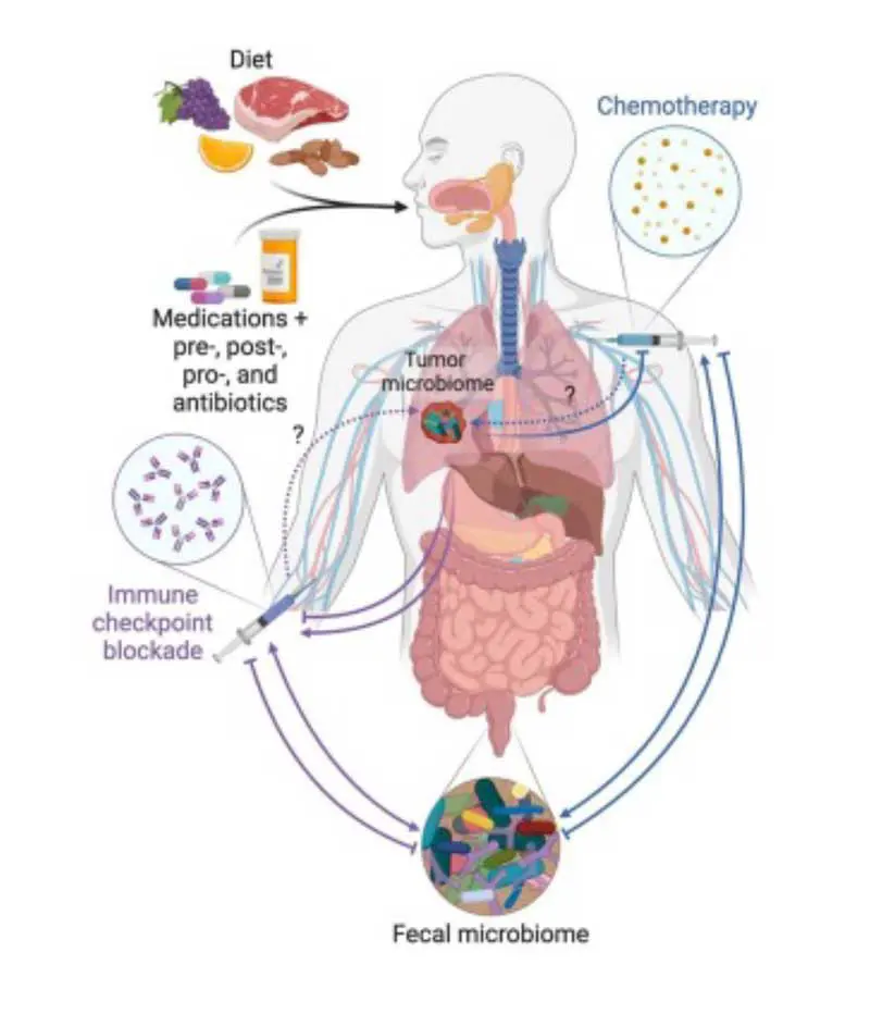
The gut microbiota is a key regulator of the TME, which is capable of driving or inhibiting cancer progression through its regulatory roles in host pathways, immune responses and metabolic processes. It is now known that cancers often have their own intra-tumour microbiome.
Helicobacter pylori is a causative agent in gastric cancer and, in 1994, was the first bacterium classified as a group 1 carcinogen by the International Agency for Research on Cancer. We now know that specific microbes have a direct impact on carcinogenesis through inflammation, genotoxicity and immune modulation.
Other examples are Fusobacterium nucleatum in colorectal cancer and Porphyromonas gingivalis in oral cancer (Li et al, 2022; Lamont et al, 2022; Akbari et al, 2024). Some strains of Escherichia coli produce colibactin, a genotoxin implicated in colorectal cancer.
F nucleatum also metabolises chemotherapeutic agents to alter their effectiveness and, in colorectal cancer, it triggers chemoresistance by reducing efficacy of 5-fluorouracil and oxaliplatin to activate autophagy pathways in cancer cells. (Chrysostomou et al, 2023; Jiang et al, 2023; LaCourse et al, 2022; Zhang et al, 2019; Galeano Niño et al, 2022).
Fungal profiling of 35 tumour types has linked species such as Candida and Malassezia with gastrointestinal cancer and pancreatic cancer, respectively. These fungi synergise with bacteria to promote cancer progression and modulate immune responses (Straussman et al, 2012; Allwood et al, 2020).
The microbiota within cancers is a complex, dynamic ecosystem, encompassing a diverse spectrum of microorganisms; for example, bacteria, fungi and viruses. Each microbial group interacts within the TME, influencing cancer initiation, progression, metastasis, and therapeutic response.
The alimentary tract mucosa is the first barrier against microbial invasion and, when compromised, provides a gateway for bacteria to infiltrate tissues and induce carcinogenesis. Periodontal disease and poor oral hygiene compromise the mucosal barrier in the oral cavity, facilitating translocation of bacteria such as F nucleatum to distant sites, including the colon, where it adheres to and invades colonic epithelial cells, promoting colorectal carcinogenesis by activating β-catenin signalling.
Streptococcus anginosus, another oral microbe, has been identified in oesophageal and liver cancer tissues (Fu et al, 2024; Morita et al, 2003).
Hypoxic cores, a defining feature of a tumour, are an ideal environment for anaerobic bacteria such as Clostridium species, which thrive in low-oxygen conditions and can promote angiogenesis and tumour growth (Baima et al, 2024; Singh et al, 2025).
The presence of microbiota-cancer associations require further investigation in veterinary patients; nevertheless, maintaining good oral health and an optimum gut microbiome through diet should be part of modern day canine and feline cancer management.
Antibiotics disrupt microbial homeostasis, leading to dysbiosis and overgrowth of pathogenic bacteria, which alter responses to cancer therapy and increases bacterial colonisation of canine and feline tumours (Curran et al, 2021).
Malignant cells are characterised by the upregulation or activation of many signalling pathways involved in proliferation, apoptosis, invasion and angiogenesis. Targeting these signalling pathways is a strategy for managing human prostate cancer (McCarty, 2004), and canine and feline cancer in the future.
Derangements in signalling are required for the formation of a fully invasive tumour, and include the following (Hanahan and Weinberg, 2000):
These can all be targeted for medical and nutritional interventions.
Cancer often results in a spectrum of malnutrition, cachexia and sarcopenia (Bossi et al, 2021).
Cancer cells reprogramme their energy pathways to support rapid growth and proliferation, and preferentially use glycolysis for energy production, even in the presence of oxygen. This process, called the Warburg effect, allows cancer cells to generate adenosine triphosphate quickly, which is essential for their rapid growth (Biocrates, 2026).
Amino acids are crucial for cancer cell metabolism and growth, and the non-essential amino acids serine, glycine and glutamine support tumour development and progression. These amino acids are vital for lipid and protein synthesis, and for generating antioxidant defences, which are essential for cancer cell proliferation and survival.
A diet restricted in serine and glycine significantly impaired tumour growth, leading to smaller tumours and increased lifespan in mice (Jiménez-Alonso et al, 2023; Chen et al, 2024; Lyu et al, 2025; Huang et al, 2025). Glutamine is a major energy source and building block for cancer cells – especially when glucose is scarce. Cancer cells increase their uptake of glutamine to meet their high metabolic demands, to synthesise nucleotides and other amino acids. Glutamine also replenishes intermediates in the Krebs cycle, ensuring a continuous supply of carbon for macromolecule biosynthesis (Cancer Today, 2022; Lv et al, 2025).
Metabolic reprogramming of branched-chain amino acids (BCAAs) – leucine, isoleucine, and valine – is another feature of cancer, and these amino acids function as signalling mediators and epigenetic modulators, facilitating tumour progression through immune evasion, and conferring resistance to therapeutic agents (He et al, 2026). This is problematic because isoleucine is also necessary to support lean muscle mass and other bodily functions (Gu et al, 2019).
Cancer cells use BCAA metabolism to fuel anabolic processes, activate oncogenic signalling cascades including mTOR and PI3K/AKT, and remodel the tumour microenvironment. Therefore, a paradoxical role exists for BCAAs, as they can modulate anti-tumour immunity, but also the development of pharmacological inhibitors targeting this pathway (He et al, 2026).
Malignant tumours produce metal-containing enzymes, matrix metalloproteinases (MMPs), and zinc-dependent MMPs degrade extracellular matrix components, facilitating tumour invasion, metastasis and angiogenesis, making them key players and potential therapeutic targets in cancer.
Gelatinases such as MMP2 and MMP9 are particularly important in degrading type IV collagen in basement membranes, facilitating endothelial cell migration and new vessel formation.
MMPs, including MMP1, MMP9, MMP10, MMP11, and MMP13, are consistently upregulated across multiple cancer types, while others such as MMP2 and MMP7 show variable expression. High MMP expression often correlates with poor prognosis and aggressive tumour behaviour, so they are pharmacologic targets with MMP inhibitors (Quintero-Fabián et al, 2019; Winer et al, 2016). Zinc-restriction might be another option.
Dietary components and other nutritional factors are associated with cancer risk. Nutrigenomics can help identify individuals with genetic susceptibilities and guide towards cancer-preventive diets.
Nutrigenetics focuses on how an individual’s gene profile influences response to dietary components. It identifies genetic variations that affect nutrient absorption, metabolism and utilisation; for example, MTHFR gene variations can impact ability to metabolise folate, affecting its roles in the body and altering dietary folate requirements.
Epigenetics is an important part of nutrigenomics and represents changes in gene expression that occur without alterations to the underlying DNA sequence. Methylation and histone modification are two common epigenetic mechanisms influenced by nutrition; for example, folate is essential for DNA methylation, and inadequate intake can lead to changes in gene expression, contributing to health issues.
Finding interactions between genes and diet in an individual can potentially help identify target molecules to prevent and/or reduce symptoms of cancer (Ardekani and Jabbari, 2009).
In humans, It is estimated that diet influences 30% to 40% of all cancer cases (Davis and Milner, 2004), and breast, prostate, liver, colon and lung cancers are linked to dietary intake (Davis, 2007).
In addition to influencing DNA methylation, bioactive components in foods such as calcium; zinc; selenium; folate; vitamins C, D and E; carotenoids; flavonoids; indoles; allyl sulphur compounds; conjugated linoleic acid; and n-3 fatty acids may influence carcinogen metabolism, cell signalling, cell cycle control, apoptosis, hormonal balance and angiogenesis (Surh, 2003; Johaning et al, 2002).
In humans, according to the World Health Organization (WHO), evidence supports:
In addition, high doses of β-carotene may increase risk of lung cancer, and oesophageal cancer is associated with obesity, probably due to acid reflux (Key et al, 2020).
No nutritional causes of cancer have yet been identified in dogs or cats; however, rations producing dysbiosis (such as raw meat diets) may contribute to translocation of microorganisms to the TME.
In numerous rodent studies, vitamin E supplementation has been shown to inhibit tumour growth (Lu et al, 2010; Li et al, 2011; Lambert et al, 2009; Ju et al, 2009; Guan et al, 2012).
However, clinical trials in humans have shown conflicting results for vitamin E with/without selenium supplementation (Mahabir et al, 2008; Helzlsouer et al, 2000; Huang et al, 2003). A meta-analysis of 14 trials concluded that “there is no convincing evidence that the use of supplemental multivitamins or any specific vitamin affects the occurrence or severity of prostate cancer” (Stratton and Godwin, 2011).
In the Women’s Health Study (https://whs.bwh.harvard.edu), vitamin E supplementation did not significantly affect the incidence of colon, lung or total cancers, and in the Physicians’ Health Study II randomised control trial (https://phs.bwh.harvard.edu), supplementation with vitamin E or vitamin C for eight years did not reduce the risk of prostate cancer or all other cancers (Lee et al, 2005; Gaziano et al, 2009).
In the Selenium and Vitamin E Cancer Prevention Trial (SELECT) involving 35,533 men, it was concluded that “dietary supplementation with vitamin E significantly increased the risk of prostate cancer among healthy men” (Lippman et al, 2009; Klein et al, 2011).
In a survey of owners of Scottish terriers, an inverse association was reported between consumption of vegetables, but not cruciferous vegetables, at least three times per week (odds ratio, 0.30; 95% confidence interval, 0.15 to 0.62) and risk of developing transition cell carcinoma of the urinary bladder (Raghavan et al, 2005). These observational study findings are in accordance with the WHO claim that vegetable intake is associated with reduced cancer risk, but further studies are needed to confirm the results and determine the mechanism.
Mammalian biosystems and aetiopathogenesis mechanisms are similar across species, and further studies are needed to confirm whether the diet-cancer associations recognised in humans and other species are also present in cats and dogs.
It is imperative to try to ensure adequate intake of energy, protein, fats, minerals and vitamins, levels of which are all greatly increased by cancer – particularly in the presence of rapidly growing malignant tumours. This often requires multiple meals a day and, if voluntary intake is insufficient, assisted feeding techniques should be employed (Davies, 2025). Pets should be weighed regularly with muscle mass assessment, and energy intake in the form of fats and proteins adjusted accordingly. Feeding a highly digestible diet with high-quality protein consisting of a high amount of essential amino acids and minimum amounts of aromatic amino acid (leucine and isoleucine) and other amino acids (serine, glycine and glutamine) is known to support tumour growth (Jiménez-Alonso et al, 2023; Chen et al, 2024; Lyu et al, 2025; Huang et al, 2025). Although cats and dog can use carbohydrates for energy, high carbohydrate diets should be avoided, as they are not essential nutrients and are preferentially used for energy by cancer cells.
In human medicine, the Enhanced Recovery After Surgery (ERAS) Society protocols in cancer surgery significantly improve recovery outcomes. These involve nutritional support to reduce malnutrition, which is common in cancer patients; avoidance of prolonged fasting and early initiation of oral feeding to support gut function and recovery. (Ljungqvist et al, 2017; https://erassociety.org/guidelines). To facilitate this in case of post-surgical anorexia, it is prudent to place feeding tubes as early as possible, such as at the time of surgery (Davies, 2025).
Cancer surgery involving the alimentary tract can have significant effects on nutrition; for example, if the distal ileum has to be excised, bile acid recycling is impaired and fat malabsorption is an expected consequence, resulting in steatorrhea and other nutrient losses.
Under such circumstances, a low-fat diet would be advisable. Chemotherapy can impact nutrition in many ways including anorexia, nausea, vomiting, mucositis, organ damage and food aversion.
Catabolic medications: corticosteroids and other medications are catabolic drugs, increasing host metabolic rate and energy requirements.
Radiotherapy: when applied to the head/neck may decrease saliva production, causing dry mucous membranes, ulceration, bleeding, soreness, reduced taste, reduced sense of smell, nausea, vomiting and diarrhoea.
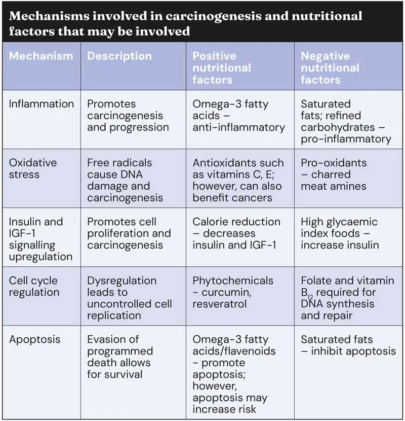
Nutrients and bioactive compounds in food can influence carcinogenesis by modulating immune status, inflammation, oxidative stress, DNA repair, cell proliferation and apoptosis. Diet also affects gut microbiome, which is a key player in cancer development and response to therapy (Ruban et al, 2025).
The following list covers some nutrients and other bioactives in foods studied with theoretical potential for clinical use in managing cancer, broken down into the nutrient; cancer; and the effect on it.
Anthocyanidins. In natural plant pigments red, purple, blue in fruits, vegetables, and flowers; non-small-cell lung cancer cell lines; synergistically inhibited growth (Kausar et al, 2012).
Anthocyanins: From tomato (prolonged life in Trp53−/− knockout mice; anti-cancer effect in vivo, restoring the function of tumour suppressors Trp53 (Butelli et al, 2008).
Antioxidants. These accelerated lung cancer progression in mice; increased cancer burden and mortality in a mouse melanoma model by activating RHO proteins; antioxidants, including catechins, quercetin, and vitamin C, accelerated the rate of cachexia progression in the host by promoting tumour growth; they could potentially reduce free radical damage; however, human clinical trials have failed to prove antioxidants are beneficial in cancer, and other studies show they are detrimental (Sayin et al, 2014; Le Gal et al, 2015; Assi et al, 2016).
Arginine. Canine lymphoma – as an adjunct to diet and medication; arginine supplementation at a rate of 2% was considered beneficial in cancer by supporting immune status (Ogilvie et al, 2000); however, arginine is important for cancer development, and arginine restriction is now considered beneficial in managing cancer (Feng et al, 2025).
β-carotene. Decreased risk for prostate, breast, oesophageal, lung, stomach, pancreas, and bladder cancer is claimed in some studies; anti-angiogenic; inhibits secretion of VEGF, expression of MMP2 and MMP9; increased expression of TIMP1 and TIMP2; and decreased tumour-directed capillaries (Aprile et al, 2015; Chen H-Y et al, 2012; Tosetti et al, 2009); however, the findings of a systemic review showed β-carotene supplementation was associated with an increased risk of lung cancer (Kordiak et al, 2022).
Black raspberries extract – phytochemicals. In vitro – effect on immune cells; decreased CD4+ and CD8+ T cells expressing the CTLA-4 and PD-1 receptors, reducing the load of T cells susceptible to immunosuppression (Mace et al, 2014).
Capsaicin from hot chilli peppers. Reportedly has antitumour properties mediated by cytotoxicity and immunological adjuvancy against at least 74 varieties of cancer; anti-angiogenic; however, it also promotes epithelial–mesenchymal transition (Tosetti et al, 2009; Luján-Méndez et al, 2023).
Copper. Prostate cancer – in vitro – promotes prostate cancer; negative effect on prostate cancer cells – increases zinc MMP expression (Parr-Sturgess et al, 2012).
Curcumin. In studies on lung cancer (mice and cell lines), hepatocellular carcinoma (human cell line), leukaemia, cervical cancer, colon cancer and breast cancer, the following positive results were reported: anti-angiogenic; inhibited lung metastasis in mice; inhibited cellular migration and invasion in a human hepatocellular carcinoma cell lines; decreased MMP9 secretion dose-dependently; inhibited invasion and metastasis by increasing E-cadherin expression in lung cancer cells; inhibited telomerase, decrease replicative immortality and genomic instability of cancer cell; caused apoptosis; improved the therapeutic efficacy of cisplatin (Mosieniak et al, 2016; Rana et al, 2015; Kazemi-Lomedasht et al, 2013; Yadav and Aggarwal, 2011; Menon et al, 1999; Lin et al, 1998; Chen et al, 2008; Tosetti et al, 2009; Baharuddin et al, 2016).
Garlic – allicin. Hepatocellular cancer; anti-carcinogenic properties that modulate cellular energetics (Chu et al, 2012).
Genistein – soy isoflavone. Human colon cancer cell lines; apoptotic induction, cell cycle arrest, anti-angiogenic, antimetastatic, anti-inflammatory (Tuli et al, 2019).
Grape seed proanthocyanidins. Skin cancer; reduced UV-induced immunosuppression, a mechanism which allows for the onset and progression of skin carcinogenesis.(Vaid et al, 2011).
Green tea catechin. This is highly toxic to starved dogs. In prostate cancer, gastric cancer and colon carcinoma, positive benefits were reported (Zhu et al, 2007; Jung al, 2001).
Iron. Bladder, lung, colon and oesophageal cancer; negative effects (Torti et al, 2018).
Lycopene. Decreased risk for prostate, breast, oesophageal, lung, stomach, pancreas, and bladder cancer; anti-angiogenic (Chen M-L et al, 2012; Aprile et al, 2015).
Probiotic – Bifidobacterium. Melanoma in mice; treatment of transverse aortic constriction mice with Bifidobacterium alone was sufficient to induce an antitumor immune response and control tumour growth (Kong et al, 2025; Matson et al, 2018).
Resveratrol – a naturally occurring polyphenolic stilbene derivative. Various, canine melanoma cells and osteosarcoma; modulated apoptosis, cell cycle regulation, inflammation, angiogenesis, and metastasis, its interaction with cancer stem cells and the tumour microenvironment; proliferation of osteosarcoma cells in vitro and in vivo was reduced by resveratrol (Kursvietiene et al, 2023; Fukuoka et al, 2023; Peng and Jiang, 2018).
Sulforaphane (SFN) – naturally occurring compound found in cruciferous vegetables. In studies on prostate cancer (mice and cell lines), lung cancer (mice), malignant glioma (human cells), non-small cell lung cancer and osteosarcoma, it was found that SFN inhibited invasion and metastasis in multiple cancer cell lines and mouse models, and it inhibited the expression and activity of telomerase (Abbas et al, 2016; Juge et al, 2007; Singh et al, 2009; Thejass and Kuttan, 2006; Bertl et al, 2006; Higdon et al, 2007; Huang et al 2012; Chen et al, 2015; Wang et al, 2004; Herman-Antosiewicz et al, 2007; Kim et al, 2011).
Vitamin D – high plasma 25-hydroxyvitamin D (25(OH)D). Colorectal cancer (humans); 25(OH)D is associated with lower risk of colorectal cancer; however, most studies do not show any benefit (Sluyter et al, 2020; National Cancer Institute, 2026).
Vitamin A – isotretinoin and etretinate. Benign cutaneous neoplasia and cutaneous lymphoma – positive in dogs; squamous cell carcinoma, papillomavirus infection – positive in humans (White et al, 1993; Ramos et al, 2022; Everts and Akuailou, 2021; Sedjo et al, 2002).
Vitamin E. Lung cancer – positive; prostate cancer – no effect or increased risk; colon cancer – no effect, positive in mice; all cancer – no effect (Mahabir et al, 2008; Lambert et al, 2009; Stratton and Godwin, 2011; Lee et al, 2005; Gaziano et al, 2009; Ju et al, 2009; Guan et al, 2012).
Vitamin E and selenium. Prostate cancer – increased risk (Klein et al, 2011).
Mixture of trans-resveratrol, quercetin, vitamin E and selenium. Colorectal cancer – positive in mice (Czapla et al, 2022).
While some nutrients and bioactives in food are worthy of further study, as can be seen from the previous list, some have conflicting evidence and, in the author’s opinion, none of these should be used unless sufficient evidence exists and clinical trials prove they are safe and more beneficial to the patient than to the cancer. As an example, apoptosis is meant to eliminate dysfunctional or pre-cancerous cells, but increased apoptosis can also cause initiation of certain types of cancers such as hepatocellular carcinoma (HCC). During the progression of HCC, this increase in the regeneration of hepatocytes leads to more genetic errors, thereby predisposing to the development of cancer.
Resveratrol exposure has been shown to induce differentiation in canine oral mucosal melanoma cells, enhancing their sensitivity to cisplatin and increasing the mRNA expression of melanoma differentiation markers, such as microphthalmia-associated transcription factor (Fukuoka et al, 2023), but randomised, controlled trials are needed.
Nutritional interventions can be used to target tumour metabolism (Chen et al, 2017):
Cancer can have a significant effect on gastrointestinal tract function due to a local effect, but also secondary effects: functional/metabolic. Cancer affects the host energy needs and results in metabolic changes long before obvious weight loss occurs, and these metabolic effects may persist for a long time after a malignancy is removed (Ogilvie et al, 2000).
Tumour cells preferentially use glucose for energy by anaerobic glycolysis and generate lactate. In dogs, this has been associated with (Ogilvie et al, 2000):
Peripheral insulin resistance may be present which has resulted in a recommendation that glucose and lactated Ringer’s IV solutions should be avoided in cancer patients. High carbohydrate diets should be avoided, and less than 20% dry matter basis is recommended (Ogilvie et al 2000).
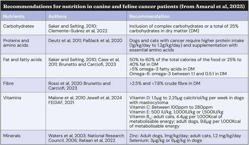
All tumours use protein at the expense of the patient for their own growth, enzyme synthesis, and in the case of endocrine neoplasia, to synthesise hormones. Some tumours preferentially use protein and amino acids for energy, and of course protein is preferentially used for energy in cats. Plasma amino acid concentrations may fall or rise in dogs with cancer, and protein intake should be higher than for healthy animals: 30% to 45% dry matter (DM) in dogs, and 40% to 50% DM in cats (Ogilvie et al, 2000).
Historically, 2% arginine supplementation has been recommended (Ogilvie et al, 2000) because of its role in supporting immunity; however, recently it has been demonstrated that arginine also supports cancer growth and is paradoxically identified as being both necessary for cancer growth and normal immune function (Albaugh et al, 2017). Arginine restriction is currently being used in managing cancers (Feng et al, 2025).
Water-soluble vitamin B complex deficiency is likely if cancer patients are polyuric, as they are in paraneoplastic syndrome due to hypercalcaemia, or associated with anti-cancer medications such as corticosteroids, and supplementation is advisable.
Weight loss can be profound in cancer patients and is due to a complex combination of reduced food intake, reduced food digestion/absorption/utilisation, increased metabolic rate, diversion of nutrients to the tumour, drug effects and radiation effects. Cytokines such as IL-1, IL-6 and TNF-α play central roles in appetite inhibition. As a result, cancer patients often have a combination of simple and stress starvation (Argilés et al, 2014; Molfino et al, 2017) and it is a major concern for pet owners (Johannes et al, 2019).
Cancer patients should be weighed regularly, and increased energy requirements must be met throughout the disease process.
Weight loss in cancer is associated with increased lipolysis, and cancer patients have altered lipid profiles such as high cholesterol, high triglycerides, and high VLDL-TG cholesterol. These lipid changes can affect immunocompetence; for example, omega-3 fatty acids are precursors to PGE3 and leukotriene LT-B5. Some tumour cells have difficulty using fats for energy, so increasing intake is not increasing energy supply to those tumours.
Omega-3 fatty acids have been shown in rodent studies to inhibit tumourigenesis and cancer spread. Eicosapentaenoic acid (EPA) decreases protein degradation, inhibits neovascularisation, reduces radiation damage to normal skin cells and has an anti-cachectic effect. Omega-3 fatty acids also reduce secretion of tumour necrosis factor, IL-1α, IL-1β and IL-2, and inhibit MMPs that are responsible for damaging adjacent healthy tissue to allow local metastatic spread (Joshi et al, 2025; Freitas and Campos, 2019; Ogilvie et al, 2000). As a result, it is recommended that fats should provide 50% to 65% of daily metabolisable energy.
It is imperative to provide increased food supply to cancer patients for maintenance, to maintain their ability to raise an immune response to fight the cancer and to prevent secondary infections. Expect to have to provide assisted feeding (Davies, 2025), so place feeding tubes as early as possible – especially when general anaesthesia is to be administered for a procedure such as imaging or surgery.
Maintaining adequate nutrition enhances quality of life, prolongs disease-free intervals, increases survival time, reduces or prevents toxicity associated with cancer therapy, ameliorates metabolic alterations that accompany cancer and can have anti-cancer effects.
Benefits of dietary support demonstrated in dogs include weight gain, better tolerance to surgery, radiotherapy, chemotherapy and better immune response: immunoglobulin and complement, white blood cell phagocytosis (Ogilvie et al, 2000).
It is commonly claimed that antioxidant supplementation is beneficial in cancer management; however, human clinical trials have failed to prove the benefits of antioxidants and some studies have shown harmful effects (Sayin et al, 2014; Le Gal et al, 2015; Wang et al, 2016) and that they may accelerate cachexia (Assi et al, 2016).
Few clinical trials involving dietary interventions in canine and feline cancer exist. In one study (Ogilvie et al, 2000), a diet formulated with low carbohydrates, high protein and high omega–3 fatty acids supplemented with arginine was given to dogs with lymphoma treated with doxorubicin chemotherapy. The dogs on the experimental diet showed significant benefits: increasing docosahexaenoic acid (DHA; C22:6) levels were significantly (P < 0.05) associated with longer disease free intervals and survival times for dogs with stage III lymphoma.
This study formed the basis for Hill’s Prescription Diet n/d, but this product has now been discontinued. The company instead recommends Prescription Diet ONC Care, which incorporates omega-3 fatty acids and a microbiome-moderating mix of nutrients (Anthony et al, 2023; Iliopoulou et al, 2013).
Restricted iodine in food (Hill’s Prescription Diet Thyroid Care y/d) is associated with resolution of signs of hyperthyroidism in clinical trials in cats with thyroid adenoma. This therapeutic pet food has an iodine content of 0.1ppm to 0.3ppm, compared to 1.5ppm to 99ppm found in other foods. Thyroid hormone concentrations decrease within three weeks and most cats are euthyroid within eight weeks. Based on clinical studies, the majority of cats are controlled if they continue eating only y/d.
Dietary recommendations have been made for canine and feline cancer patients (Amaral et al, 2025); however, some of these recommendations (for example, the authors’ recommendation to use carbohydrate as an energy source) are in conflict with other studies, and a lack of clinical trials exist to support them.
The following studies suggest potential benefits of some nutritional supplements, but they have not been reported in randomised controlled clinical trials:
Nutritional intake is an important factor in the initiation, maintenance and management of cancer in dogs and cats.
Further studies are needed to establish optimal levels of different nutrients and bioactives for preventing and managing different types of cancer; however, the following recommendations can be made.
Mike Davies qualified from the RVC, has RCVS postgraduate certificates in veterinary radiology and small animal orthopaedics, and holds a fellowship by examination in clinical nutrition in cats and dogs. He is an RCVS specialist in veterinary nutrition (small animal clinical nutrition). Mike has worked in academia and private practice, and for several pet food manufacturers and pharmaceutical companies. He speaks internationally on clinical nutrition and geriatrics, and founded the original City and Guilds certificate in small animal nutrition, and the BVNA certificates in small animal and exotic nutrition. He runs Provet’s certificate course in clinical nutrition.