
20 Jul 2026
Ellie Barker RVN discusses four issues often seen in practice and how nurses can help to manage them.

Image: Tanya / Adobe Stock
Eye conditions in companion animals are commonly encountered in general practice and their presentation can range from mild irritation to serious vision-threatening disease.
Early recognition and appropriate management are essential to prevent complications such as chronic discomfort, vision impairment or loss of the eye.
This article outlines four commonly encountered eye conditions, how veterinary nurses can help manage them and key points to consider for effective client communication.
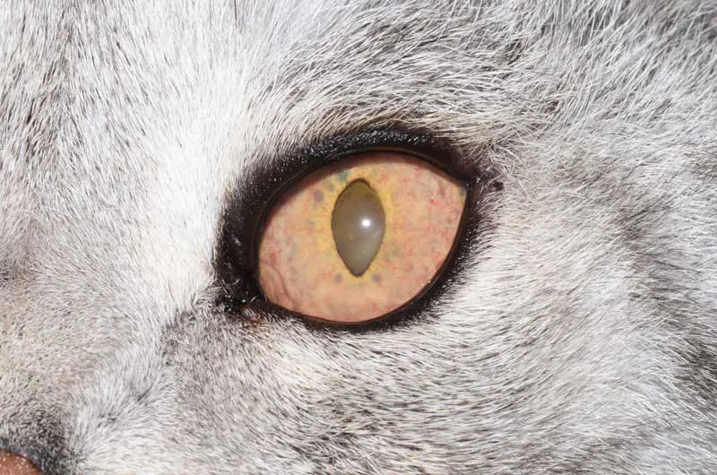
Uveitis refers to inflammation of the uveal tract, a part of the eye comprised of the iris, ciliary body and choroid. It is a potentially serious condition that can lead to vision-threatening complications if not treated promptly. Patients typically present with blepharospasm (squinting), conjunctival hyperaemia (redness of the conjunctiva), corneal oedema (cloudiness of the cornea) and miosis (constricted pupils, Figure 1).
Although uveitis can affect any cat or dog, certain breeds can be predisposed, for example, golden retrievers. Definitive diagnosis of the underlying problem can be challenging as there are a wide range of causes, including primary ocular causes (for example, corneal ulcers, cataracts, ocular trauma) and secondary systemic disease (for example, infectious or inflammatory diseases, and neoplasia). In some cases, an underlying cause is not identified, and these cases are referred to as “idiopathic”.
A diagnostic work up by the vet requires a thorough general physical and ocular exam, including measurement of IOP (intraocular pressure). Uveitis is typically associated with a low IOP, helping to distinguish it from glaucoma, which presents with a high IOP. Ophthalmic examination with a slit lamp, blood testing to check for infectious/systemic disease, and further imaging (for example chest x-rays and abdominal ultrasound) to detect potential neoplasia may be indicated to aid diagnosis in the absence of a primary ocular problem.
Treatment generally involves a combination of topical and systemic NSAIDs (non-steroidal anti-inflammatory drugs) or corticosteroids, to relieve discomfort and reduce inflammation, as well as specific treatment of any underlying systemic cause.
Nursing considerations include preventing self-trauma by placing an Elizabethan collar (E-collar), monitoring IOP, fluorescein staining the corneal surface, minimising stress as this can exacerbate clinical signs, and supporting repeat exams and ongoing investigations. Care should be taken to avoid any pressure around the neck when performing IOP measurements as this can give falsely elevated readings.
Clear communication with owners is essential to manage their expectations, as frequent follow-up visits will be necessary. If left untreated, uveitis can progress to glaucoma, a painful condition with increased ocular pressure that can lead to reduced vision or blindness, eventually necessitating removal of the globe in most cases.
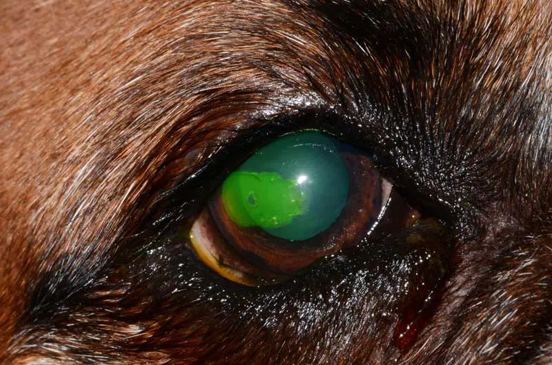
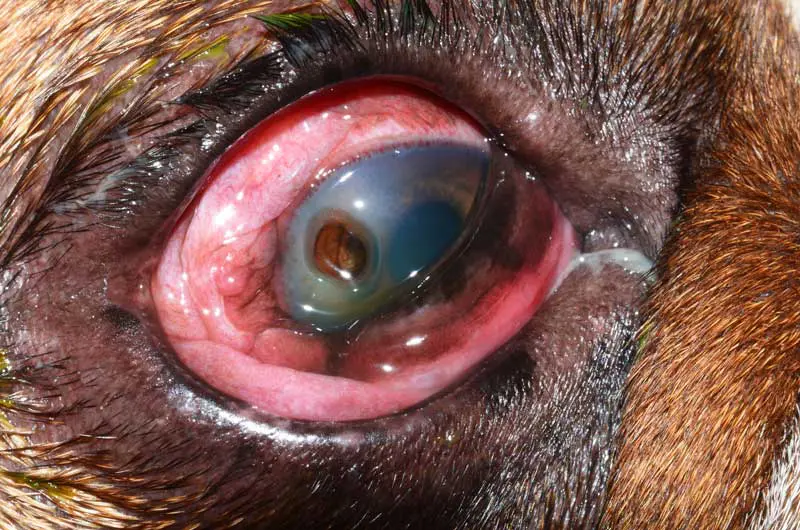
Corneal ulcers are a common presentation in dogs and cats. They are painful and vary in severity, ranging from superficial (Figure 2) to deep (Figure 3) or perforating lesions. Clinical signs include blepharospasm, conjunctival hyperaemia, ocular discharge and, occasionally, a cloudy appearance of the cornea.
Corneal ulcers typically develop secondary to trauma; however, underlying conditions such as keratoconjunctivitis sicca (KCS; “dry eye”) or entropion (rolling in of the eyelids) may predispose some patients. Brachycephalic breeds whose eyes are more prominent and have poor corneal innervation and sensation are also at increased risk. Feline herpes virus (FHV-1) is a common cause of corneal ulcers in cats associated with signs of keratitis and infection.
Diagnosis is typically made using a fluorescein dye that will stain the area of ulceration bright green. It is important to flush the ocular surface with sterile saline afterwards as the stain can cause local irritation. If available in the practice, the vet can use a slit lamp to assess the depth and severity of the ulcer.
Treatment depends on the type of ulcer. Superficial ulcers are generally managed with symptomatic, empirical topical antibiotic eye drops in addition to oral medication such as pain relief. They typically carry an excellent prognosis. Chronic superficial ulcers may require a diamond burr debridement, while more severe or infected ulcers often require intensive medical management and/or surgical intervention.
Nursing involvement can vary depending on the severity but generally includes administering topical and oral medications, fitting an E-collar to prevent self-trauma, and monitoring for any changes suggestive of deterioration (for example, increased ocular discharge and signs of worsening ocular discomfort). Hospitalisation may be necessary for intensive medical management, requiring five minute intervals between application of different eye drops to the same eye to enable effective drug absorption.
Monitoring of the corneal ulcer by repeated fluorescein staining at follow-up appointments and comparison to the previously ulcerated area can also be performed by registered veterinary nurses.
Client education, particularly demonstrating correct eye drop application and reinforcing the importance of rechecks, even if the eye looks better or appears more comfortable, can help to minimise complications. Failure to treat appropriately can lead to deterioration of the ulcer and potentially perforation of the cornea.
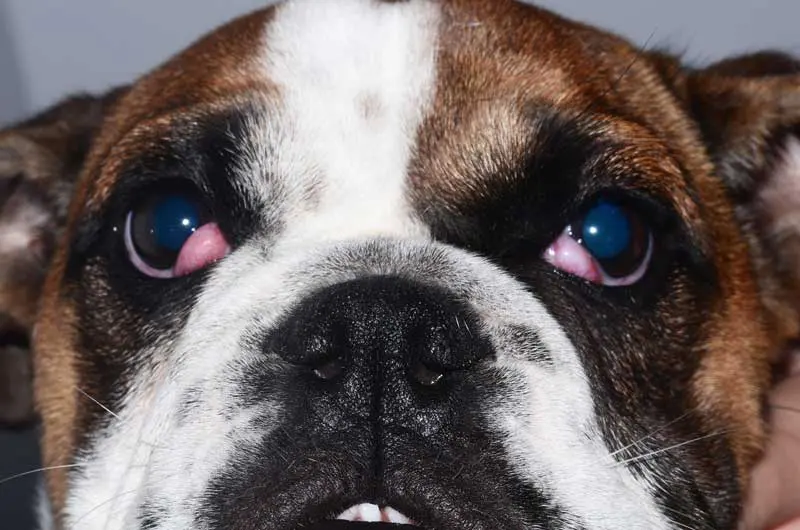
Cherry eye (Figure 4) refers to a prolapse of the third eyelid gland. It presents as a red-pink mass at the inner corner of the eye. It is most commonly seen in younger dogs and is an infrequent condition in cats. In the early stages, the gland may prolapse intermittently and can be gently massaged back in place but will often become permanently proptosed with time.
Initial management involves keeping the exposed gland moist with regular lubricating eye drops and reducing inflammation with prescribed anti-inflammatory medications. Surgical replacement of the gland back into its normal position is required and recommended for a long-term resolution. Recovery from surgery usually takes around 10 days, during which time exercise should be restricted and the E-collar worn to prevent patient interference. Removal of the gland is not recommended as this can lead to decreased tear production, increased risk of dry eye disease and corneal ulcers.
Nursing care involves preventing self trauma by fitting an E-collar, demonstrating how to administer prescribed medications to owners, and educating owners on how to monitor for potential complications such as signs of infection.
If left untreated, cherry eye can lead to several other conditions that can be painful and irritating to the patient such as conjunctivitis, infection, ulceration of the conjunctiva and the gland, KCS and corneal ulceration.
Conjunctivitis refers to inflammation of the conjunctiva, the pink tissue lining the sclera (white part of the globe) and inner aspects of the eyelids. It typically presents with conjunctival hyperaemia, ocular discharge – which may be clear, white, yellow, or green – and blepharospasm. Both eyes are often involved and concurrent respiratory (mostly in cats) and dermatological (more frequently in dogs) signs may be present.
Conjunctivitis has numerous causes. In cats, feline herpesvirus is the most common cause, while in young and middle-aged adult dogs, it is often secondary to bacterial infections, allergies, or environmental irritants. In middle-aged to older dogs, KCS is the most common diagnosis. Less frequently conjunctivitis may be associated with eyelid abnormalities, nasolacrimal duct obstruction or other ocular diseases.
Diagnosis will depend on the clinical history, examination findings and further testing, if indicated. Diagnostic tests include Schirmer tear testing, fluorescein staining, IOP measurements and, in some cases, conjunctival culture and sensitivity. More complex cases may require nasolacrimal duct flushing, allergy testing or conjunctival biopsy.
Treatment typically involves promoting local hygiene with sterile saline washes, topical medications – which may include antibiotics and ocular lubrication – and sometimes systemic medication is also required. When conjunctivitis is secondary, addressing the underlying cause is essential for effective case management.
Nursing responsibilities may include assisting with and performing diagnostic tests, administering treatment, educating owners on correct application of prescribed medications, and how to clean and flush the ocular surface.
Client communication is particularly important in cases of infectious disease. For example, bacterial conjunctivitis in dogs can be contagious to other dogs in the same household, while feline herpes virus is also highly contagious and can spread through direct and indirect contact with other cats in the home. Owners should be advised on isolation, strict hygiene, and stress reduction in multi-cat households.
Owners should be made aware that some conditions such a KCS or allergies will require long-term or lifelong management, and while symptoms often improve within a few days, completing the full course of prescribed treatment is essential to prevent recurrence.
Veterinary nurses can play a vital role in the management of ocular disease, from early recognition and diagnostic support, to patient care and client education. Immediate escalation to the vet is warranted if any of the following are observed:
Understanding when to escalate, how to monitor, and supporting owners through treatment plans will help to ensure the best possible outcome for each individual patient.
Fact sheets about eye conditions, referral information, advice request, CPD and the Vet Eye Care blog can be found at https://veteyecare.co.uk/