12 Nov 2021
Megan Brashear discusses how an appropriate fluid therapy plan is an important element of a successful anaesthetic event, and the steps teams can take both before and during procedures to ensure a smooth experience.
Megan Brashear
Job Title
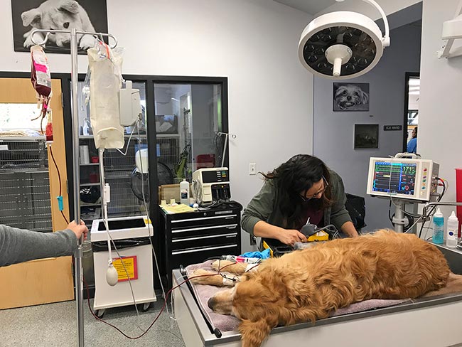
Anaesthesia monitoring.
In emergency veterinary medicine, the team cannot always plan for the perfect time to take a patient to surgery.
While the ideal anaesthetic patient is one that is hydrated, stable and pain-free, emergency situations often arise that require surgery and general anaesthesia in less than ideal situations.
Haemoabdomen, gastric dilatation-volvulus, urethral obstruction, septic abdominal effusion and major wounds may require an unstable patient to be anaesthetised. Gastrointestinal obstruction and minor wound care may allow for a longer patient stabilisation period, but still require patient preparation and close monitoring.
While the perfect situation cannot always be created, veterinary nursing teams can take steps to ensure anaesthesia runs as smoothly as possible, even with a critical patient.
Appropriate fluid therapy prior to and during anaesthesia can make a significant difference in the anaesthetic experience, and should have close and careful attention paid prior to anaesthesia induction.
The goals of preoperative fluid therapy are to optimise patient tissue perfusion, balance oxygen delivery and correct any electrolyte abnormalities (Boller and Boller, 2015).
Knowing that the effects of both IV and inhaled anaesthetics can worsen perfusion, it is important to perform fluid resuscitation prior to anaesthetic induction.
The veterinary nursing team must first perform a physical examination on any patient – and in this population of critical patients, pay careful attention to the physical signs of hypovolaemic shock.
In dogs, as water and electrolytes are lost (through vomiting, diarrhoea or blood loss from trauma) or not maintained (inappropriate consumption due to illness or injury), they will begin to show clinical signs of this loss.
As their body attempts to compensate for decreased volume, early decompensatory shock will present as tachycardia, snappy or bounding pulse quality, pale mucous membranes, prolonged capillary refill time, decreased mentation and hypotension.
Recognition and understanding of these clinical signs are important, as shock must be addressed prior to general anaesthesia.
Once identified, hypovolaemic shock is addressed with fluid therapy, which requires careful monitoring by the veterinary nursing staff. Prior to beginning any course of fluid therapy treatment, patients must be assessed for heart disease, as cardiogenic shock is treated and managed differently.
In severe cases of hypovolaemic shock, multiple rounds of bolus therapy may be required to ensure proper tissue perfusion. In this acute therapy stage, a balanced crystalloid is administered to replace water and sodium. These isotonic crystalloids will only remain in the intravascular space for about 30 minutes, requiring their rapid administration prior to anaesthesia (Boller and Boller, 2015). The blood volume of dogs is 90ml/kg and cats is 60ml/kg.
While few patients require replacement of an entire blood volume, this number is used to then calculate appropriate fluid bolus amounts.
In dogs with moderate fluid losses and minor vital sign abnormalities (such as minor wound repairs and early gastrointestinal disease), it is appropriate to administer a 10ml/kg to 15ml/kg fluid bolus. In more severe cases of fluid loss and dramatic changes to patient vital signs, 20ml/kg to 30ml/kg may be required, again administering as a fast IV bolus (Boller and Boller, 2015).
In patients with gastrointestinal disease, it is common to see an increase in diarrhoea and/or vomiting after IV fluid therapy. The veterinary nursing team should note these ongoing losses and expect to replace them with an increase to the fluid therapy plan.
Cats can be a challenge, as the physical signs of hypovolaemic shock in cats are not always straightforward, nor do they mirror the same signs as dogs.
Cats are often bradycardic or even present with a normal heart rate when in hypovolaemic shock. However, they are often hypotensive and hypothermic, allowing the team to rely on blood pressure and body temperature as clinical signs (Chalifoux et al, 2021).
Fluid resuscitation in cats requires more caution, as cats can more easily suffer from fluid overload. Administering not enough fluids to restore perfusion – or too much fluid, leading to pulmonary oedema – is a fine line in emergency feline patients. In these cases, it is important for the veterinary nursing team to perform frequent physical exams and monitor for respiratory changes.
Colloids encompass a class of IV fluids made up of larger molecules compared to crystalloid fluids.
Colloids can be further divided into synthetic (various brands of starch fluids) and natural (blood products such as packed red blood cells and plasma).
Because of these large molecules, colloids will remain in the intravascular space longer than crystalloids, and provide improved circulating volume and perfusion (Byers, 2017).
Over the past 10 years, synthetic colloids have come under scrutiny in both human and veterinary medicine as their use has not been proven to improve patient outcomes and may be linked to acute kidney injury in critically ill human patients (Cazzolli and Prittie, 2015).
Concern exists for similar outcomes in veterinary patients – and while synthetic colloids are still used, teams should be aware of the potential risks.
During this phase of preoperative fluid resuscitation, the veterinary nursing team plays a vital role in monitoring and giving real‑time feedback on the patient’s status.
Perfusion improvement can be monitored by frequent measurement of heart rate (dogs should decrease as they improve perfusion; cats may see an increase in heart rate), mucous membrane colour, capillary refill time and mentation. These values should be reported to the veterinarian at least after each fluid bolus as the results will be used to determine next steps.
Aside from monitoring heart rate, mucous membrane colour and capillary refill time, blood pressure measurement is an important piece in monitoring critical patients receiving fluid therapy. Veterinary nurses should note that the blood pressure value means little without use of other monitoring parameters, meaning a normal blood pressure in a dog still experiencing significant tachycardia does not mean all is well.
Shock index can be calculated by dividing the patient’s heart rate by its systolic blood pressure, and can be a good tool to use and track patient improvement during fluid therapy. As the patient improves, the shock index will decrease (Boller and Boller, 2015).
Lactate is also a valuable tool that can be used to determine the success of fluid therapy.
Lactate is produced when the cellular energy requirements are greater than the aerobic energy production. When oxygen delivery to the tissues is decreased due to hypovolaemic shock, cells cannot produce adequate amounts of energy and lactate levels will increase.
The actual patient lactate value is of less importance than the trend of lactate in response to fluid therapy, so serial measurements should be performed to appropriately interpret lactate.
Even in patients that present to the hospital with extremely elevated lactate levels, if those levels decrease in response to therapy, that patient has a good chance for recovery (Boller and Boller, 2015).
A good goal when using serial lactate measurements to track fluid therapy effectiveness is to see a 50% decrease in lactate every one to two hours.
Veterinary nurses are now monitoring IV fluid rates, heart rate, mucous membrane colour, capillary refill time, blood pressure, shock index and lactate, but other important parameters need to be monitored in these patients prior to anaesthesia induction.
Urine output and urine specific gravity can give further clues to fluid therapy success – and depending on the length of time from fluids to urine, can be significant.
In a patient that has received large volumes of crystalloid therapy, yet produced no urine over a period of hours, this information must be followed up. Gentle palpation or cage‑side ultrasound can be used to determine bladder size and if further investigation is needed.
Patients receiving large volumes of fluids should also be monitored for fluid overload, consisting of closely watching respiratory rate and effort, ausculting lung sounds, and watching for the formation of peripheral oedema.
Remembering that the goal is to have the best possible candidate for general anaesthesia, these additional monitoring parameters must have attention paid (Boller and Boller, 2015).
Significant trauma adds an additional layer of necessary thought when deciding on a fluid therapy plan prior to anaesthesia. Trauma patients can suffer from massive haemorrhage requiring immediate volume replacement.
Hypertonic saline may be administered to facilitate a quick draw of fluids from the interstitial to the intravascular space. Hypertonic saline has a much higher sodium concentration (usually 7% sodium chloride) compared to physiologic saline (0.9%), which draws fluid into the intravascular space in an attempt to equalise the sodium content. Relatively small volumes are needed (5ml/kg) administered over 5 to 10 minutes.
The intravascular volume expanding effects of hypertonic saline last about 30 minutes, but their administration may keep the patient alive long enough to allow a large bolus of crystalloid fluids or other therapies time to work.
The fluid shift that occurs with the use of hypertonic saline can help to reduce intracranial pressure in head trauma patients, as well as modulate inflammation.
Hypertonic saline should not be used as the sole fluid used for resuscitation as it can cause dramatic changes in the patient’s serum sodium levels (Byers, 2017).
Coagulopathy in trauma patients is not a new concept and can be the result of loss of clotting factors due to bleeding, dysfunction of clotting factors, or haemodilution of clotting factors resulting from fluid resuscitation.
The “triad of death” in trauma – coagulopathy, hypothermia, and acidosis resulting from haemorrhage – is a critical combination that can quickly live up to its namesake and cause mortality in trauma patients. This triad can be seen in polytrauma patients and contributes to a more severe disease process.
The key is quick recognition of these factors and mitigating the effects on the patient.
Knowing that trauma patients that receive large quantities of crystalloid fluids can suffer from haemodilution, consideration for blood products (such as fresh frozen plasma) may be considered early in the treatment process, even prior to the animal showing signs of uncontrolled haemorrhage.
Acidosis can result from hypoperfusion and elevated lactate, respiratory malfunction leading to respiratory acidosis, and decreased cardiac output. Acidosis serves to compound hypocoagulability in trauma patients.
We know that hypothermia decreases platelet function and white blood cell function, decreases cardiac output, decreases oxygen delivery to tissues, and causes a decreased response to catecholamines.
Trauma patients must be kept warm, and veterinary nurses should consider covering and warming normothermic trauma patients as they present to the hospital, knowing that treatment with fluids and pain management can reduce body temperature (Palmer, 2017).
Preparing critical patients for general anaesthesia and surgery requires a team effort with everyone doing their part to examine, treat, monitor and be ready for changes in the patient status.
While an appropriate fluid therapy plan is not the only part to this plan, it is an important key to a successful anaesthetic event.
Megan Brashear
Job Title