
1 Sept 2026
RVNs: integral in investigation and management of otitis externa
Deborah James RVN explains how VNs are best placed to help with the ongoing care of this disease and to ensure client compliance.

Deborah James
Job Title
Image: charnsitr / Adobe Stock
- This article from VN Times (September/October 2026) can count towards your CPD – 20 minutes
Otitis externa is one of the most common dermatological problems encountered in veterinary practice. While veterinary surgeons are responsible for diagnosing and deciding the best course of treatment action, veterinary nurses play an integral part in the investigation and management of the disease, alongside educating clients to ensure compliance.
What is otitis?
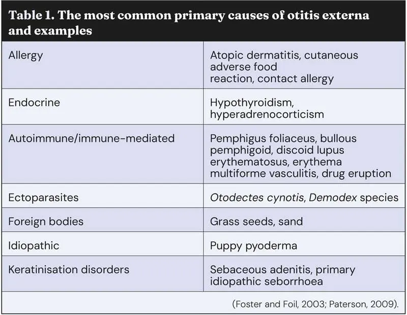
Otitis, or otitis externa, is the acute or chronic inflammation of the external ear canal and is seen in around 10% to 20% of dogs, and 2% to 6% of cats, in practice (Maginn, 2016). As with any skin problem, a full comprehensive history, physical and otic examination must take place prior to further diagnostic tests (Paterson, 2021). The aetiology and pathogenesis of otitis externa can be divided into four parts using the acronym PPSP: primary causes, predisposing factors, secondary infection and perpetuating factors.
Causes of otitis
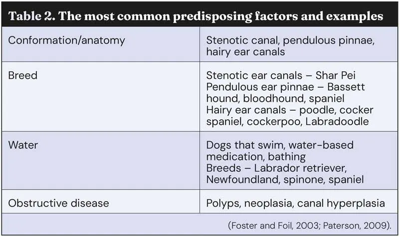
Primary causes are generally present in every case of otitis externa, and skin problems are largely responsible for starting the inflammatory process in the ear (Paterson, 2021; Table 1). Predisposing factors are those which contribute to the disease itself but do not cause any inflammation, although it is important to note overlap can occur on occasions; for example, obstructive diseases (Paterson, 2021; Table 2).
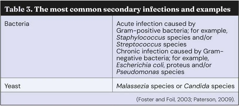
It is important to note that it is secondary infections, not infection. Infections are never a primary problem, but rather a secondary one that develops due to inflammation by primary causes in combination with perpetuating and predisposing factors – normally of a yeast or bacterial origin (Paterson, 2021). These infections require resolution, but only once the other factors have been treated (Table 3).
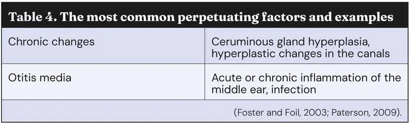
Finally, perpetuating factors are those that drive an already established disease process and are generally the most important reason for the long-standing disease. Without treatment to resolution, the otitis will relapse (Paterson, 2021; Table 4).
Veterinary nurse role
Veterinary nurses play a vital role in identifying primary causes to helping manage secondary infections and predisposing factors. Alongside this, veterinary nurses can assist by taking samples from the ears and performing microscopy and identifying yeast and/or bacteria, ensuring a prompt and correct treatment for the patient while utilising key nursing skills.
Not only this, but veterinary nurses play a role in owner compliance by demonstrating how to apply the medication to their pet and supporting them through the process, for less able clients, offering a daily consult for medication to be applied.
Sampling and microscopy
Sampling is fairly quick and simple, with the equipment being readily available and, the majority of the time, in-house assessment will deliver a definitive diagnosis leading to quicker treatments, so they are more cost effective for the client.
Sampling should be performed for every patient presenting with otitis, and it is essential for follow-up appointments to be booked to ensure that the treatment has been effective.
As previously mentioned, otitis externa can occur due to a number of causes and, ideally, if the patient allows it, otoscopy should be performed prior to sampling for a multitude of reasons (Miller et al, 2013):
- To check for a foreign body.
- To check the integrity of the tympanic membrane.
- To check for erythema, ulcers, polyps – a friable ear canal can cause trauma and even haemorrhage.
To conduct sampling, the following equipment should be assembled:
- Microscope slides (frosted ends enables ease of labelling, but any microscope slide will do).
- Coverslip.
- Liquid paraffin.
- Cotton buds.
- Pencil.
- Immersion oil.
- DiffQuik.
- Microscope.
After preparing your equipment, the patient should be restrained. A veterinary nurse can be the restraint or the sample taker, and even both. Restraint can be the difference between collecting a sample or not, and minimising head movement is key in this process. Following this, an impression smear can be taken. Normally, a cotton bud can be used to collect the sample, but in cases where the patient is head shy, it can sometimes be easier to gently insert a gloved finger and collect a sample that way and transfer it on to a microscope slide (Forsythe, 2016).
In addition to this, both ears should be sampled, as the findings between the ears may differ. Unfortunately, not every case is gold standard. Patients may be in too much pain or too head shy as a result of chronic ear problems that ear sampling while conscious would be impossible. Sedation may be required for these patients to allow a thorough examination or, in some cases, where the ear canal is so stenotic, medications such as steroids can be administered to reduce the inflammation and swelling. As a result, this opens the ear canal, which in turn would improve medication penetration and provide pain relief to the patient (Hensel and Hensel, 2021).
Once the sample has been taken, gently roll the cotton bud over the slide, or if the sample was collected using a gloved finger, then press and roll the finger on to the slide, being careful not to smear the sample (Macfarlane, 2017).
Label the sample, remembering to label which slide is from which ear. Allow the smear to dry and then stain the slide using DiffQuik.
If the sample is purulent, then dip the slide 5 to 10 times in each stain and rinse gently with water at the end (Toma et al, 2006). If the sample is waxy, then miss out the blue fixative stain, as a risk that this might dissolve the sample exists. Instead, use 5 to 10 dips in the red (eosinophilic) and purple (basophilic) stain before rinsing gently with water (Toma et al, 2006). It is not necessary to heat fix these slides; however, give the slide time to dry or, if you do not, gently blot any excess liquid away (Macfarlane, 2017).
Once the slide is dry, apply a small drop of liquid paraffin or immersion oil on to the sample and cover with a coverslip – microscopes are designed to look through a fluid layer, so this way the image will be clearer and the microscope will remain clean and free from contamination (Macfarlane, 2017). If the sample is suspected to contain ectoparasites, then apply a drop of liquid paraffin to the slide. To transfer the sample, roll the cotton bud in the liquid paraffin, then apply a coverslip and examine. All ectoparasites are visible at ×4 magnification, but ×10 may be required to determine if it is alive (Macfarlane, 2017). If the slide is being examined for cytology only, then ×4 or ×10 initially can be used to identify an area of interest.
Once an area has been located, the magnification should be increased to ×40 or ×100 to identify yeasts, bacteria and inflammatory cells (Shaw, 2016).
Conclusion
Veterinary nurses are more than capable of assessing the external ear canal, both macroscopically and cytologically, to help the veterinary surgeon to make a diagnosis.
Client-facing nurse communications can help with the administration of therapy, provide owner support during treatment to increase compliance and help with follow-up assessments. The veterinary nurse can improve patient care, increase the speed of recovery and, through owner education, reduce the chance of reoccurrence.
- This article appeared in VN Times (September/October 2026), Volume 26, Issue 9/10, Pages 14-17
Deborah James qualified from Hartpury College in 2017 with a BSc in veterinary nursing science and works as team leader at Vet’s Klinic. She has completed an ISFM certificate in feline nursing and is in her second year of her MSc in veterinary nursing at the University of Glasgow. She has two cats at home: Tipsy, a 10-year-old hand-reared cat, and six-year-old Frank. In her spare time, you can find Deborah snuggled with a book or teaching and participating in pole fitness and aerial arts.
References
- Forsythe PJ (2016). Acute otitis externa: the successful first-opinion ear consultation, In Practice 38(S2): 2-6.
- Foster A and Foil C (2003). BSAVA Manual of Small Animal Dermatology (2nd edn), BSAVA, Gloucester.
- Hensel P and Hensel N (2021). Treating otitis externa in dogs, Today’s Veterinary Practice, https://todaysveterinarypractice.com/dermatology/treating-otitis-externa-in-dogs
- Macfarlane C (2017). How to perform ear sampling and undertake microscopy, The Veterinary Nurse 8(5): 272-276.
- Maginn K (2016). Management of otitis externa and the veterinary nurse’s role, The Veterinary Nurse 7(1): 25-32.
- May ER (2026). Image gallery: normal vs abnormal ear cytology, Clinician’s Brief, www.cliniciansbrief.com/article/image-gallery-ear-cytology
- Miller WH et al (2013). Diseases of eyelids, claws, anal sacs, and ears. In Miller WH et al (eds), Muller and Kirk’s Small Animal Dermatology (7th edn), Elsevier, St. Louis: 724-773.
- Papajeski B (2020). Ear cytology: sampling, processing, and microscopic evaluation, Today’s Veterinary Nurse, https://todaysveterinarynurse.com/clinical-pathology/veterinary-ear-cytology
- Paterson S (2009). Otitis scares me - where do I start? Veterinary Ireland Journal 6: 316-317.
- Paterson S (2021). Otitis externa: the role of the veterinary nurse, The Veterinary Nurse 12(7): 306-310.
- Shaw S (2016). Pathogens in otitis externa: diagnostic techniques to identify secondary causes of ear disease, In Practice 38(S2): 12-16.
- Toma S et al (2006). Comparison of 4 fixation and staining methods for the cytologic evaluation of ear canals with clinical evidence of ceruminous otitis externa, Veterinary Clinical Pathology 35(2): 194-198.