
14 Jul 2026
Kieran Borgeat BSc, BVSc, MVetMed, CertVC, DipACVIM, DipECVIM-CA, FHEA, FRCVS and Joshua Hannabuss BVetMed, MVetMed, DipACVIM, MRCVS discuss perspectives for the UK in 2026 on this common problem in older canines.
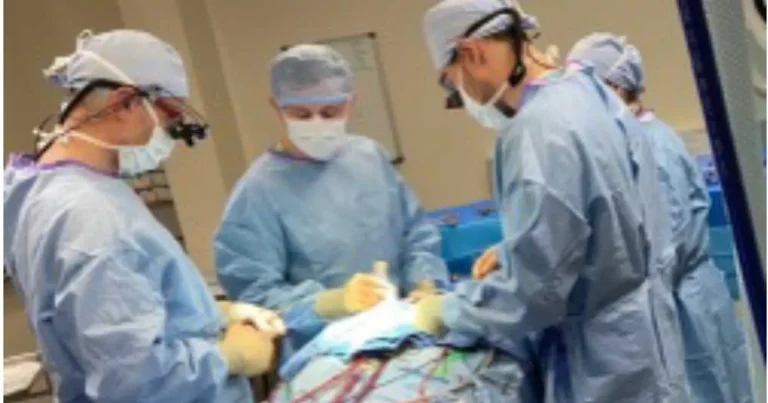
Figure 1. Open heart surgery for primary mitral valve repair requires a large team of surgeons, scrub nurses, anaesthetists and perfusionists, alongside a heart-lung machine for cardiopulmonary bypass and facilities for cardioplegia.
Mitral valve disease (MVD) is a huge contributor to ill health in older dogs. Estimates of prevalence differ according to the population reported, but up to 3.5% of all dogs in the UK are probably affected, based on primary-care records from the VetCompass database (Mattin et al, 2015).
In smaller breeds (dogs with less than 20kg bodyweight), the overall risk is doubled compared to larger dogs, and particular breeds are more at-risk, including the cavalier King Charles spaniel, whippet, Yorkshire terrier, Chihuahua and bichon frise. Larger breeds, such as the Labrador retriever and the German shepherd dog, are also affected.
The impact of MVD on animal welfare is huge, with an associated burden on owner mental health through caregiver burden associated with long-term medication. Demographic data obtained pre-pandemic (McMillan et al, 2024) estimates a population of 1.2m dogs in the UK from predisposed small pedigree breeds, with at least an additional 160,000 dogs of various cross-breeds that are considered to be at-risk (such as the cavapoo or cavachon). Just under half of dogs were reported to be in the senior or geriatric category.
Imagine that just 20% of these 1.4m dogs were to become affected by MVD over their natural lifespan, and one in three of these dogs goes on to develop severe mitral regurgitation which leads to a recommendation for medical therapy. We would then have more than 93,000 dogs on treatment over the next 5 to 10 years.
Although they serve to illustrate the potential impact of MVD on pets and their owners in the UK, these numbers may well be an underestimate of post-pandemic dog ownership and, therefore, under-represent disease burden.
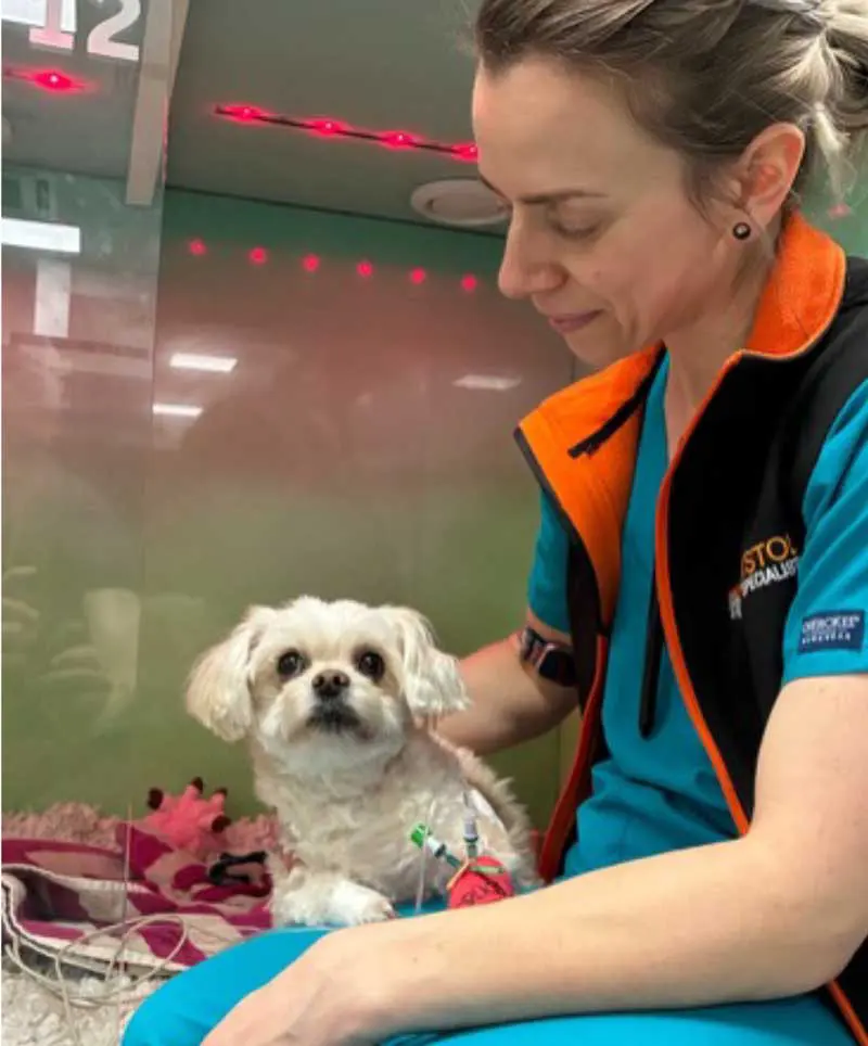
From large disease registries, we can see that the natural history of MVD is generally benign, with approximately 70% of affected dogs dying for non-cardiac reasons (Borgarelli, LOOKMitral registry, personal communication, 2024). To identify the one in three dogs with MVD that do progress, echocardiographic measurements provide the best predictive value. Early detection of cardiac remodelling in dogs with MVD is vital, because treatment with pimobendan can prolong the asymptomatic stage of disease and reduce risk of heart failure every day by approximately one-third, prior to the onset of overt clinical signs (Boswood et al, 2016). Although some data suggests that pre-clinical treatment with benazepril and spironolactone can help maintain reduced cardiac chamber size for longer, there is no effect on time to the development of heart failure signs (Borgarelli et al, 2020).
Once dogs reach the stage of developing congestive heart failure signs (increased respiratory rate and effort), average survival time is reported at 9 to 12 months (Häggström et al, 2008; Beaumier et al, 2018; Brložnik et al, 2023), with individual variation either side of this median. Two-year survival rate is approximately 15%.
A large case series from a single centre in the US (Carlson and Stern, 2026) suggests a potential improvement in survival for dogs with heart failure treated off-licence with Entresto, a combination drug comprising valsartan (an angiotensin-II receptor blocker) and sacubitril (which reduces breakdown of B-type natriuretic peptide). Although enough to justify further study, this publication does not evaluate safety or efficacy in a controlled trial, so a treatment recommendation cannot be made based on this. However, many owner-facing online forums recommend this drug highly, so enquiries from owners have become frequent in recent months, and it is worth practising vets being aware of the publication and public interest. We have prescribed Entresto for dogs when they have established heart failure signs that have become refractory to standard medical therapy, requiring more than 6mg/kg/day furosemide to control pulmonary oedema. Owing to the lack of rigorous scientific data, there is no consensus amongst cardiologists on the utility of this drug in dogs.
Left heart chamber size (increased diameter of the left atrium and left ventricle, suggesting greater neurohormonal activation and volume retention) and increased Doppler-derived mitral inflow velocity (suggesting worse mitral regurgitation volume) are straightforward echo measurements that can be used to spot the worst cases of MVD in dogs.
More challenging echocardiographic measurements can be used to obtain estimates of mitral regurgitant fraction, defined as how much of total stroke volume goes backwards during systole (with more than 50% considered severe). This is useful to identify the dogs with the most severe regurgitation prior to secondary remodelling of the heart.
Different cardiologists take different approaches to this, ranging from volumetric techniques or surrogate markers, but a high level of echo experience and training is required to ensure reliability, owing to compound error in the formulae used for these calculations.
A simplified composite score of echo variables, the MINE 2 score (Vezzosi et al 2025), may be a straightforward way to help stratify risk, and dogs with a severe MINE score have been shown to have a shorter survival time than those with moderate or mild scores, separate to the prognostic valve of MVD disease stage based on the American College of Veterinary Internal Medicine Consensus Guidelines (Keene et al, 2019).
In recent years, assessment of mitral regurgitation severity beyond chamber size has become more important, to make decisions around surgical intervention for MVD before cardiac remodelling or clinical signs are so advanced that procedural risk becomes too great.
After a decade of regular caseload, the RVC remains the UK’s only centre with an active programme for mitral valve repair at open heart surgery, under cardiopulmonary bypass. It has demonstrated success in overcoming the steep learning curve for bypass surgery in dogs with close support from transfusion medicine, anaesthesia, cardiology and emergency and critical care services. The actual repair technique has been refined through collaborating with Masami Uechi of the Jasmine Animal Referral Hospital in Tokyo – a world pioneer for canine mitral repair surgery.
The RVC team schedules two cases most weeks. The procedure allows inspection and repair of the mitral valve in a bloodless field, through cessation and protection of the heart with cardioplegia, and circulatory support with a heart-lung machine. The valve is repaired through the placement of artificial chordae tendineae and an annuloplasty in addition to potential repair of other defects (such as valve clefts).
Consumable cost is relatively low, but the expertise and team size required is high. Postoperative care can range from relatively low levels of ICU care with additional medical therapy (anti-arrhythmic medication), to requirement for more intensive support including mechanical ventilation, and temporary pacing (rare).
The reported results for surgical valve repair are incredibly good overall, and the Jasmine team have reported 97% three-year survival after surgery. The RVC team’s documented survival to discharge rate of 80% to 90% (Brockman et al, 2025; Palacios Jimenez et al, 2026), and many cases have no significant mitral regurgitation after surgery. The cost of surgery is £24,200 as a package, after diagnosis and determination of candidacy, but for the majority of dogs this route offers an effective cure for their disease, coming off all medical treatment and with negligible risk of cardiac death after successful discharge from hospital.
Because cardiopulmonary bypass does pose a significant risk factor, and the cost of open heart surgery may be prohibitive, options for minimally invasive procedures to reduce mitral regurgitation severity have been developed, mimicking approaches that are becoming more widely adopted in humans to perform beating-heart surgery.
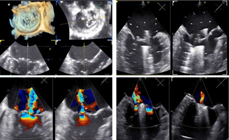
In humans, the MitraClip (Abbott Cardiovascular) and PASCAL (Edwards Lifesciences)devices have been used for more than a decade to reduce mitral regurgitation severity by decreasing valve regurgitant orifice area in patients that were considered unsuitable for bypass.
Applied to the valve through a transvascular approach and reliant on accurate advanced imaging for guidance, transcatheter edge-to-edge repair (TEER) was initially shown to be associated with a lower risk profile than bypass, but was also reported to be less effective than open heart surgical repair. Over time, refinements to the device design and the techniques of surgical teams have led to great improvements in procedural success. A significant reduction in mitral regurgitation volume and good long-term outcomes are commonplace for experienced teams in the NHS. The high cost of devices used in people served to limit access to minimally invasive mitral repair for many countries outside of Europe or the US.
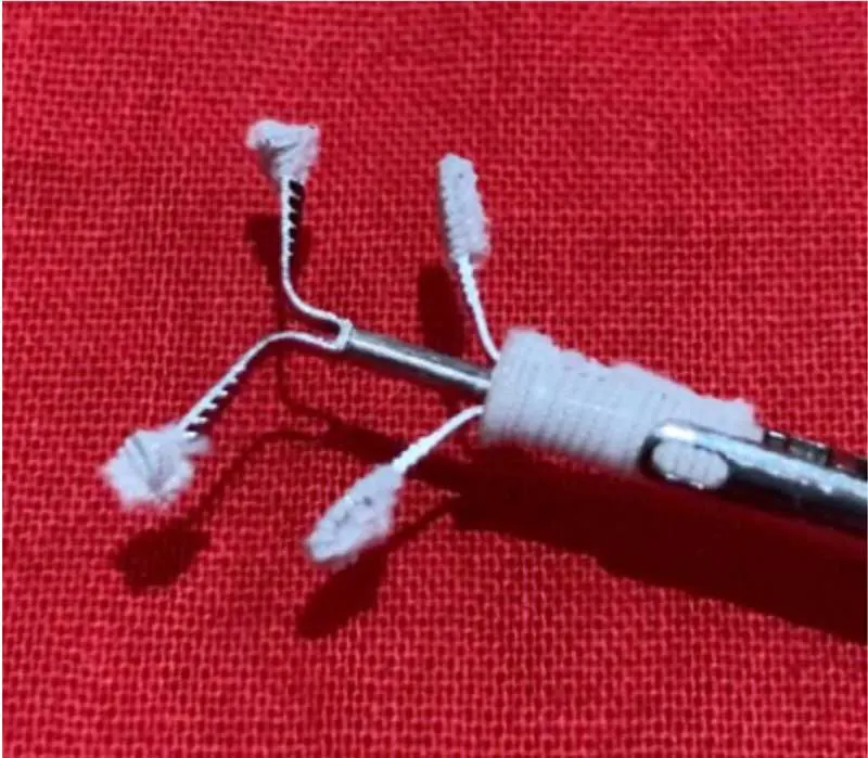
Other techniques have been developed which represent a more cost-effective option. One notable device, the V-clamp, was developed for dogs, from a device in humans which is applied to the valve using a hybrid, transapical approach. Device cost is around £5,200, compared to approximately £35,000 for the PASCAL device, so the potential for use in veterinary medicine is more realistic.
In dogs, TEER is performed through an introducer sheath placed in the left ventricular apex, on the beating heart, after exposure by a small thoracotomy. Transoesophageal echo guides the clamp placement, and position can be adjusted to maximise the reduction in mitral regurgitation. Just as in humans, some dogs benefit from a second clamp placement to adequately reduce the severity of the leak.
Outcome data from Colorado State University supports a low rate of complications: 5% to 6% device-related adverse events perioperatively (Potter et al, 2025), although most were fatal. Recovery is rapid, and most dogs can be discharged on day two after surgery, with minimal need for intensive care. Dogs can return to moderate exercise in the weeks after surgery, with restoration of full exercise regime by six to eight weeks for most.
Around 40% of dogs can have diuretic treatment stopped post-surgery, but almost all continue pimobendan therapy. Overall, two-year survival post-TEER for dogs with severe mitral regurgitation is approximately 80%, with no cardiac-related deaths reported for those dogs where TEER reduced mitral regurgitation fraction to less than 30%.
At the time of writing, we are in the early days of our TEER experience in the UK. Bristol Vet Specialists and the RVC have committed to a collaborative effort to support mutual learning of good TEER technique, to maximise successful outcomes for dogs undergoing the procedure in the UK.
After 20 cases, all but two have survived the surgery with a good reduction in mitral regurgitation severity. Regurgitant fraction was reduced to less than 30% in 13 out of 18 dogs. Sadly, 2 out of 20 did not survive because of device-related adverse events. Morbidity and mortality review has identified key learnings which have been actioned into procedural checklists, to minimise risk of repeating the same circumstances in future cases.
Mitral TEER surgery is priced around £12,000 as a package fee – so it remains high-cost, but offers an alternative to open heart surgery which may be more accessible for some families.
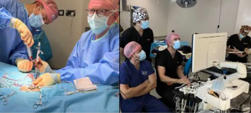
Surgical mitral repair and TEER both have a higher risk for dogs with refractory heart failure signs (stage D MVD), and both come with high-end financial implications for owners.
A lower cost alternative, which may help as part of palliative care to reduce the diuretic dose needed and help improve quality of life and longevity in dogs with advanced heart failure signs, is left atrial decompression (LAD). This transvenous procedure is performed to create an atrial septal defect and, therefore, a pressure off-let valve.
The technique is reported using trans-septal needle puncture, followed by a cutting balloon procedure, under 3D echo and fluoroscopic guidance. Novel devices are in production to prevent the defect from healing closed, as this has proven to be a common occurrence some months after surgery.
The goal of LAD is to reduce left atrial pressure at the expense of the systemic venous pressure. Around one in five dogs undergoing the procedure are reported to develop right-sided heart failure signs (ascites), although pulmonary oedema reduces in severity. Significant reduction in diuretic requirements are reported for many dogs, with a survival time of months to years (Allen et al, 2021).
Experience remains relatively limited among specialist cardiac centres in the UK, although more cardiologists with interventional experience are likely to have access to the requisite expertise and facilities needed for LAD than for TEER or bypass.
The price is significantly lower than surgical repair options, at around £5,300, and procedural risk is lower in experienced hands than open heart surgery or TEER.
However, it should be considered a palliative procedure as part of management rather than a procedure that will restore a normal lifespan in most dogs, and healing of the septostomy site seems to be a common limiting factor in sustained response.
In 2026, the UK’s pet owners and dog population with severe MVD have a greater range of treatment available to provide sustained good quality of life beyond the boundaries of classic medical management, from palliative LAD to TEER and surgical valve repair.
Awareness of the options for affected dogs, and accurate assessment of mitral regurgitation severity have both become a key part of looking after dogs with mitral valve disease.
Kieran Borgeat qualified from the University of Bristol and worked in primary care practice before undertaking a cardiology residency. He became a Diplomate in 2014 and has worked in private referral and academic practice since then. Kieran holds American and European board certification, and is an RCVS-recognised Specialist in Veterinary Cardiology. He is head of the cardiology service at Bristol Vet Specialists, and is producer and co-host of The Animal Heartbeat podcast.
Joshua Hannabuss qualified from the RVC and worked in primary care practice prior to undertaking a rotating internship at Langford Vets, followed by a cardiology internship at Anderson Moores Veterinary Specialists. He returned to the RVC for residency training, and became a diplomate of the American College of Veterinary Internal Medicine in 2024. He is lecturer in cardiology at the RVC and head of the clinical cardiology service.