8 Dec 2020
Peter Kukadia describes how to approach remote consultations to achieve a diagnosis and treatment plan for this issue.
Peter Kukadia
Job Title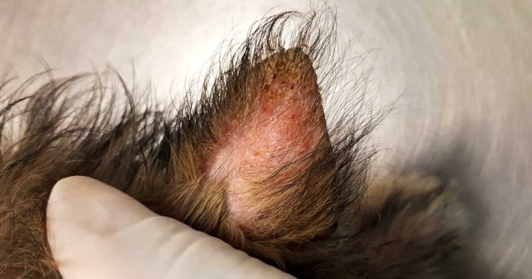
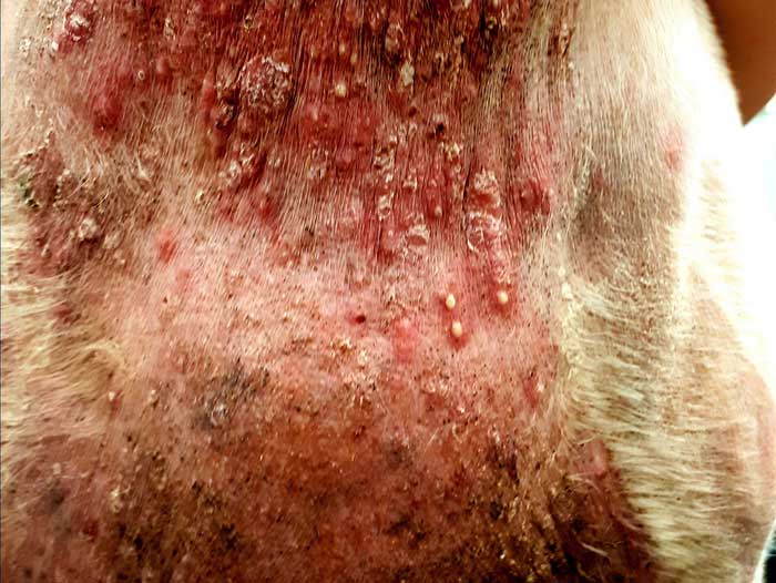
Figure 1a. Sarcoptes scabei infestation causing intense pruritus on the ear. Erythema, scaling and pinpoint crusts are present towards the apex of the pinna. Scaling is a visible accumulation of corneocytes that can be caused by inflammation. Crusting is the accumulation of serum, pus or blood that subsequently dries. In this case, the papules have ruptured to cause crusts.
In the midst of the global COVID‑19 pandemic, veterinary practices have had to steer their staff, clients and pets through a convoluted path towards offering and receiving a high standard of veterinary care.
The ability of veterinary surgeons to prescribe POM‑V drugs in the remote setting has been facilitated by the temporary relaxation of prescribing rules by the RCVS during the early stages of the pandemic in 2020. This situation is ideal for those patients that are the under the ongoing care of a veterinary practice and where a recent hands‑on physical examination supports the remote prescription of an authorised POM-V drug.
Conversely, situations exist whereby patients are under the ongoing care of a veterinary practice, but have not been examined recently to enable the safe, remote prescription of POM-V drugs. The patient may present with signs of a new, previously undiagnosed condition or a flare‑up of a pre‑existing condition.
This frustration may potentially be augmented as some owners will be unable to attend the practice premises due to self‑isolation or shielding during the COVID‑19 pandemic – and a delay in a pet receiving treatment can have deleterious effects on animal welfare and, consequently, fragment the human‑animal bond.
Dermatological diseases in companion animals will manifest in various ways, and the clue to a diagnosis lies in careful history taking and a detailed examination.
From a remote consulting perspective, clients will often be very descriptive of the lesions they are seeing, but forgo the salient historical features that often define a disease – such as pruritus seen with flea bite hypersensitivity and canine atopic dermatitis – and the omission of vital information can potentially lead to misdiagnoses.
Various modes of communication are available to practitioners. Images are able to depict skin lesion morphology, their distribution and severity of the presentation, while video recordings are able to capture valuable historical information that may aid in generating a differential diagnosis – for example, a patient seen to be scratching, rubbing, shaking, biting or over-grooming may alert the practitioner to an underlying pruritic disorder.
Through a series of dialogues, this article aims to demonstrate an approach to the remote dermatological consultation with a view to making a working diagnosis and treatment plan. Moreover, with the advent of high‑definition photography, practitioners are likely to be presented with a wide range of detailed clinical presentations – and knowledge of the types of skin lesions that occur, and their distribution in different diseases, can help towards generating a differential diagnosis list.
Practitioners are reminded, as per the current RCVS advice (at the time of writing; October 2020), they should ensure any consent given by the client is fully informed, that they (the veterinary surgeon) make detailed notes of their decision and the reasons for it, and that any decision made can be justified. Readers are advised to adhere to RCVS advice at all times when considering the remote prescription of POM-V drugs. For more information, visit https://bit.ly/3epHrBk
Ideally, a full general and dermatological history should be taken. However, due to the time constraints in general practice, the aim should be to obtain a history that is clear, concise and relevant to the pet’s situation.
Pruritus was the most common presenting sign in one study, accounting for between 30% and 40% of all dermatological consultations in general practice (Hill et al, 2006).
Many manifestations of pruritus exist, with scratching, licking, biting, chewing, rolling, rubbing and shaking being common features.
It is useful to obtain details of the time of onset and duration of the pruritus, level of pruritus over time, its relation to the appearance of any skin lesions (for example, before or after any skin lesions), seasonality, last ectoparasitic or endoparasitic treatments, and whether any in-contact pets and humans are affected, as this may suggest a contagion and zoonosis, respectively.
Primary lesions are those that arise directly as a result of the underlying pathology in the skin. Examples include macules, papules, pustules, nodules and primary, non-inflammatory alopecia.
Secondary lesions are those that have progressed from a primary lesion or occurred due to the patients’ activity – with two examples being epidermal collarettes and self‑induced alopecia, respectively. Other secondary lesions can include crusting and scaling (Figure 1a), alopecia (self-induced), hyperpigmentation and lichenification (Figure 1b).

Mrs Smith has remotely presented Rebel, a four‑year‑old male entire cross‑breed dog, with a history of skin lesions that have been present for the past five weeks:
“From what I can make out, there are a few red spots and some dandruff on my dog’s back. It has not bothered him too much initially – with an occasional bite or lick of the area – but now he has been licking his leg and his sides a lot, and has made his belly red and sore.”
From the history, it becomes apparent the licking she describes is indicative of pruritic behaviour that started off as mild, but has now progressed to the point at which the dog is pruritic for most of the day. No other animals are in contact and no humans are affected.
The owner was unsure about seasonality as this is the first time it has occurred. The dog was last dosed with an ectoparasiticide containing selamectin four months prior to presenting.
Mr Smith, who is listening in the background, also happens to state that he picked off and squeezed what looked like a flea from the dog’s back last week, but since then he has not seen anything else.
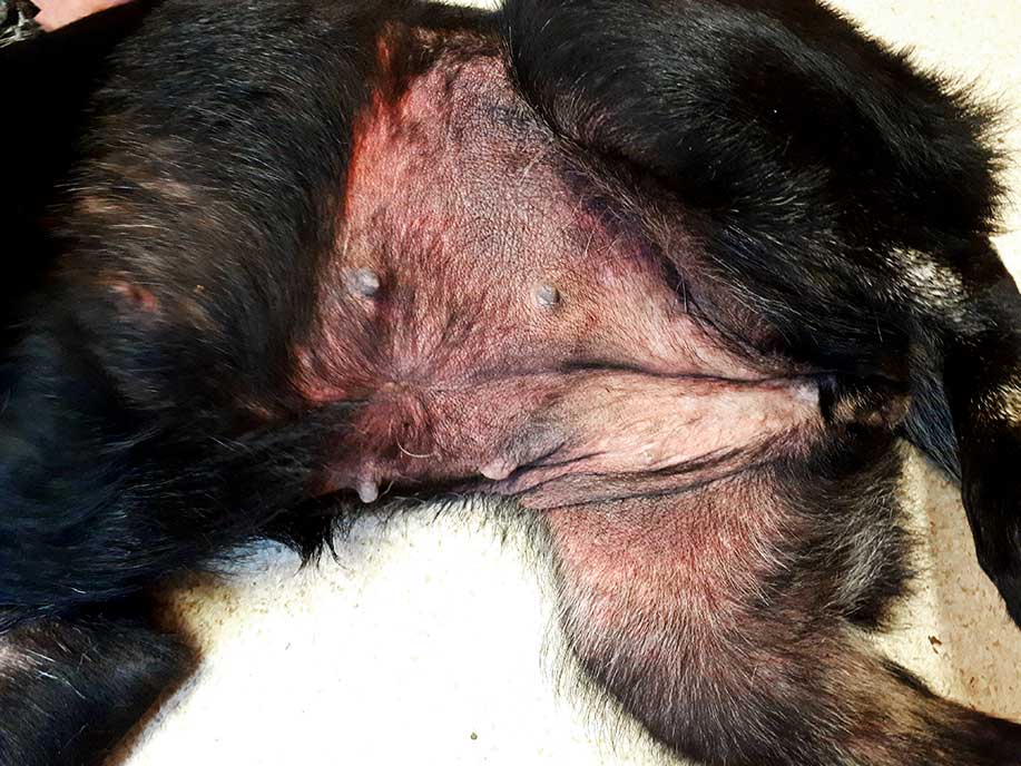
Mrs Smith has emailed two images for you to examine (Figure 2).
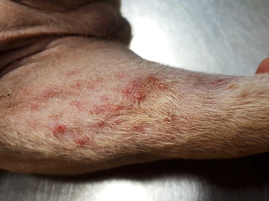
Epidermal collarettes can exist as focal lesions or can coalesce. The alopecia accompanying these lesions is often the first concern to be raised by the owner, with the scaling being subtle in some cases. The latter may be overlooked during a remote examination and it is suggested to closely examine the leading edge of these lesions as this may aid recognition (Figure 2b).
The differential diagnosis for epidermal collarette‑like lesions at first glance includes bacterial folliculitis, demodicosis and dermatophytosis.
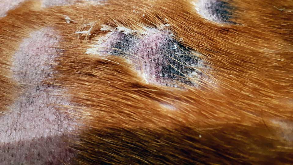
Pustular eruptions can be seen in pyoderma, demodicosis (Figure 3) and pemphigus foliaceus, with the latter being relatively rare in daily first opinion practice.
Pustules are one of several lesions that arise with superficial bacterial folliculitis (SBF), a pyoderma that affects the superficial portion of the hair follicle and the epidermis.
Other lesions that may accompany pustules are papules and secondary lesions, such as epidermal collarettes, crusting, scaling and self‑induced alopecia.
A working diagnosis of SBF secondary to flea allergic dermatitis was made. Treatment was started with the ectoparasiticide sarolaner at a dose of 2mg/kg orally, and the SBF was trial treated with a POM-V 2% chlorhexidine (CHX) and 2% miconazole shampoo twice‑weekly (with a 10-minute contact/lathering time with each treatment). After two weeks, the dog was re-examined, and the owner reported a noticeable reduction in the level of pruritus and severity of lesions. Shampooing was continued for a further three weeks, with the owner reporting some residual scale and minimal pruritus by the end of this period. Sarolaner was repeated on a monthly basis.
In the remote setting, ectoparasites cannot always be ruled out from the history alone. Therefore, the author recommended to commence an ectoparasitic treatment trial alongside shampoo therapy.
Sarolaner, an isoxazoline agent, is licensed in the UK as a POM-V drug for the treatment of fleas and demodicosis.
The patient was under the ongoing care of the veterinary surgeon at this particular practice, and after discussion with the owner, it was agreed to prescribe the two POM-V drugs as previously mentioned.
Demodicosis is included as a differential diagnosis for pustules and epidermal collarettes, with Demodex canis being the most common mite of dogs (Mueller et al, 2020). This ectoparasitosis has a localised and generalised form, with immunosuppression implicated in the pathogenesis.
Factors that may favour mite proliferation in the young are being underweight, malnourished and debilitated (Mueller et al, 2012), with an underlying immunosuppressive disease often being reportedly the cause for the adult onset form.
Canine demodicosis is primarily non‑pruritic, and patients can present with erythema, non‑pruritic alopecia/hypotrichosis, follicular casting, comedones, scaling and hyperpigmentation. Follicular papules and pustules can be present in those with more severe disease, and the presence of these two primary lesions on images should alert the clinician to include demodicosis in the differential diagnosis.
Dermatophytosis is a superficial fungal skin disease of dogs and cats, with the most common causative organism being Microsporum canis. The clinical signs can be alopecia, erythema, scaling, crusting, papules and hyperpigmentation, with pruritus generally being minimal to absent (Moriello et al, 2017).
SBF has no age, breed or sex predilection. The most common causative organism is the Gram‑positive commensal bacterium Staphylococcus pseudintermedius, which overgrows secondarily to an underlying cause that disrupts the skin’s defences; ectoparasites, allergic skin disease and endocrinopathies are examples.
The condition is variably pruritic, and in dogs the lesions may typically be on the ventral abdomen, flanks and dorsum.
In the clinic setting, a diagnosis of bacterial folliculitis is supported by clinical history, physical examination and cytology findings, with the presence of intracellular phagocytosed bacteria (most commonly cocci) within degenerative neutrophils (Beco et al, 2013a). The latter is seen more readily in surface and superficial infections.
In the remote setting, however, cytology of the lesions will not be possible – in this case, based on all the supportive evidence available, a working diagnosis of SBF was made.
The first‑line approach to treating SBF is no different in the remote setting to that adopted in the consult room.
With the ever-present scourge of antibiotic resistance, such as that seen with meticillin‑resistant staphylococci in humans and animals, the use of topical antiseptic therapy in place of systemic antibiotic therapy is both desirable and recommended (Morris et al, 2017; Hillier et al, 2014).
Various antiseptic formulations are authorised for veterinary use in the UK. CHX – in a 4% shampoo formulation – has been shown to resolve pyoderma and pruritus scores in dogs when used twice weekly for at least four weeks, with its efficacy being comparable to a course of amoxicillin-clavulanate (Borio et al, 2015).
At a concentration of 3%, Loeffler et al (2011) also demonstrated a reduction in lesions and pruritus after three weeks’ use when compared to a benzoyl peroxide shampoo.
Studies have also confirmed the antibacterial activity of CHX in different formulations. A 2% and 3% concentration in a mousse preparation was found to be active against S pseudintermedius, with the latter providing residual antibacterial activity for up to 14 days on canine hair (Ramos et al, 2019).
Furthermore, CHX impregnated on to wipes has been shown to have activity against meticillin‑sensitive and resistant S pseudintermedius, Escherichia coli and Malassezia pachydermatis (Rafferty et al, 2019).
Fortunately, several CHX products are available on the UK market that can be used safely and be dispensed without the need for a veterinary prescription. A 4% CHX shampoo – along with various mousse and wipe products – are obtainable via internet pharmacies and veterinary practices.
The prescription of systemic antibiotics in both the remote and hospital setting is not without risks to the patient and client. These include the occurrence of adverse effects to the patient, the risk to the owner handling the drug (most commonly the owner may have a penicillin allergy) and, in broader terms, the risk of further selecting for antibiotic resistance.
In view of this, it is the author’s opinion that a physical examination should be performed in the clinic setting to mitigate the risks.
Furthermore, a dermatological examination will enable practitioners to identify and sample skin lesions to rule in/out differential diagnoses to the presentation. Examples include deep skin scrapings to rule out demodicosis, and skin cytology to confirm the presence of bacteria and yeasts, such as that seen in pyoderma and Malassezia dermatitis, respectively.
Systemic antibiotics have been suggested to be used in cases with a moderate to severe generalised superficial pyoderma or deep pyoderma, where topical therapy may not be possible (due to patient and client factors), and where evidence from culture and susceptibility testing (CST) justifies their use in a particular situation (Bajwa, 2016; Beco et al, 2013b; Coyner, 2012).
It is beyond the scope of this article to discuss the indications for CST; readers are directed to an excellent article by Beco et al (2013a) for a more detailed discussion.
In the event of remotely prescribing antibiotics, consideration must be given to the likely pathogen, duration of therapy, dosage required, patient and client compliance, and any recommendations made from practice and regional prescribing protocols. More specifically, the antibiotic should be narrow‑spectrum or broad‑spectrum, with established anti‑staphylococcal activity (Beco et al, 2013b). Suitable first‑line empirical choices are shown in Table 1.
| Table 1. The recommendations for choosing specific antibiotics when treating superficial bacterial folliculitis, based on guidelines published from the International Society for Companion Animal Infectious Diseases antimicrobial guidelines working group (Hillier et al, 2014) | ||
|---|---|---|
| First-line (empirical choice) | Second-line (based on CST evidence) | Third-line (reserved for human use) |
| • Cephalexin • Amoxicillin-clavulanate • Clindamycin • Trimethoprim-sulfonamide • Cefovecina (based on Beco et al, 2013b) |
• Doxycycline • Enrofloxacin • Marbofloxacin • Pradofloxacin • Aminoglycosides (gentamicin, amikacin) • Cefovecin |
• Linezolid • Vancomycin • Teicoplanin |
| CST = culture and susceptibility testing. a Cefovecin is a third-generation cephalosporin and generally considered to be a second-line antibiotic. In situations where medicating will be difficult and/or compliance will be poor, it can be used as a first-line (Beco et al, 2013b). |
||
Second‑line antibiotics are agents that are important in both human and animal health. These should only be prescribed in situations where it is not appropriate to use empirical first‑line drugs and topical therapy (Hiller et al, 2014). Evidence should exist from a CST result that justifies their use.
Practitioners are not expected nor advised to prescribe these agents in the remote setting without a full in-clinic assessment, including the relevant diagnostic tests.
Third‑line drugs are important in the treatment of multidrug‑resistant infections in human health care and should not be used. If at this stage the case becomes challenging to manage, practitioners are strongly encouraged to consult with a veterinary dermatologist.
Pyoderma is a secondary phenomenon and will have a primary underlying cause. It is imperative to communicate to owners that without identifying an underlying cause, the pyoderma may potentially be recurrent and/or poorly resolved following initial treatment, and, as such, further investigations may be needed.
In the remote setting, general practitioners should be mindful of the various historical and physical features of some common underlying causes (Table 2), as it may become apparent during the course of the consultation as to what may have potentially triggered the onset of infection.
| Table 2. Examples of the underlying causes of pyoderma | |||
|---|---|---|---|
| Underlying cause | Salient historical/clinical features | Can it be managed remotely? | Alternative treatment options other than POM-V drugs (for dogs) |
| Ectoparasites | |||
| Fleasa | • Pruritus/irritated behaviour. • Contagious – other pets may be affected and owners may be bitten. • Ventrum, medial thighs, face, radius/ulna, tibia/tarsus (Bruet et al, 2012). |
Yes | • Fipronil • Fipronil/(s-)methoprene • Imidacloprid • Nitenpyram • Indoxacarbb |
| Sarcoptic mangea | • Intense pruritus. • Contagious – other pets may be affected, zoonosis. • Erythema, vesiculopapular eruptions, crusts, scales‑pinnae, head, elbows, tarsi, ventrum (Pin et al, 2006). |
Yesc | Noned |
| Demodicosisa | Focal/multifocal to generalised non‑pruritic alopecia, erythema, scaling, comedones, hyperpigmentation (Mueller et al, 2020). | Yesc | • Noned • Mild localised disease will resolve spontaneously in some cases |
| Allergic | |||
| Flea bite hypersensitivity | • Pruritus. • Other pets may be affected and owners may report being bitten. • Dorsolumbar area and tail (Bruet et al, 2012). Erythema, papular crusting, scaling, self‑induced alopecia. |
Yes | • Fipronil • Fipronil/(s-)methoprene • Imidacloprid • Nitenpyram • Indoxacarbb |
| Canine atopic dermatitis Diagnosis by exclusion |
• Hallmark sign: pruritus. • Erythema, papules, self‑induced alopecia, excoriations, lichenification, hyperpigmentation. • Head/concave aspects of pinnae, axillae, ventrum, inguinal areas, perineum, distal extremities (Hensel et al, 2015). |
Yese (Multifactorial disease necessitating in-clinic visits for a thorough initial work‑up) |
• Essential fatty acids • Antihistamines • Shampoo therapy • Antimicrobial wipes • Allergen avoidance |
| Cutaneous adverse food reaction | Signs almost identical to canine atopic dermatitis | Yese (Multifactorial disease necessitating in-clinic visits for a thorough initial work-up) |
• Hydrolysed diet • Home-cooked diet (Hensel et al, 2015) |
| Endocrine | |||
| Hyperadrenocorticism | • Polyuria/polydipsia, polyphagia • Bilaterally symmetrical alopecia (non-pruritic), seborrhoea, comedones, striae, calcinosis cutis (Reeder, 2013) |
No (Initial diagnosis and rechecks require blood sampling) |
None |
| Owners may incidentally report the historical features of these diseases during a remote consultation. The clinical features may also be noted on images/videos sent in by owners. The list is not exhaustive. | |||
| a The most practical method for ruling out ectoparasites from a remote perspective is to perform a treatment trial. These are three examples of the ectoparasites that present in general practice. Readers are referred to standard dermatology texts for a more comprehensive list. | |||
| b This list is not exhaustive and represents a proportion of the products available in the UK. Readers are advised to check the legal category of a medicine prior to dispensing. | |||
| c It is the author’s opinion that sarcoptic and demodectic mange can be managed successfully in the remote setting using POM-V drugs. The remote prescription of POM-V drugs should be in line with current RCVS guidelines on remote prescribing. It is the veterinary surgeon’s responsibility to ensure any prescription issued is in accordance with the veterinary medicines cascade. | |||
| d Based on a database search at www.noahcompendium.co.uk/datasheet | |||
| e Canine atopic dermatitis and cutaneous adverse food reaction are complex, multifactorial diseases that require a thorough in-clinic work-up. It is possible to manage the acute flare-ups of these conditions remotely, albeit on a short-term basis. | |||
The COVID-19 pandemic has presented many challenges to veterinary staff, clients and their pets. The majority of dermatology cases may not be routinely presented as emergencies in general practice, but they do adversely affect the quality of life of pets and their owners.
As more pet owners self-isolate due to the pandemic, the more reliance is placed on the provision of good‑quality remote veterinary care.
Dermatological problems can be acute or chronic in nature, and by applying basic consulting and visual skills in the remote consultation it is possible to manage some cases successfully for a period of time, pending an examination in the clinic.
The author would like to thank Catriona MacKinnon BVMS, GPCert(SAM), MRCVS, for her constructive criticisms during the preparation of this article.