7 Jun 2015
Kerry Hall
Job Title
Figure 7. Patient hospitalised with indwelling urinary catheter and closed collection set.
Feline lower urinary tract disease is commonly seen in practice. Patients may show chronic clinical signs or present to the clinic with acute urethral obstruction and require immediate, lifesaving medical interventions.
The RVN plays a vital role in assisting with the emergency case, but also in advising cat owners on multimodal environmental modifications, which can play a role in decreasing the recurrence.
Feline lower urinary tract disease (FLUTD) is a general term used to describe the various causes of lower urinary tract signs in cats. Common causes include urolithiasis, bacterial infection, urethral plugs or neoplasia.
Feline urological syndrome (FUS) is a term often used interchangeably with FLUTD. Feline idiopathic cystitis (FIC; also known as feline interstitial cystitis) is the most common cause of FLUTD and is diagnosed by a process of exclusion, where no specific underlying cause can be identified. FLUTD or FUS is considered one of the most common diagnoses in cats, affecting 1% annually in the UK (Gunn-Moore, 2003).
As a general rule, FLUTD is more commonly seen in:
Clinical signs of FLUTD include:
Emergency cases of urethral obstruction (UO) will often display the same signs, but will be unable to pass urine. As this progresses, the patient will become more distressed. On clinical examination, the tip of the penis may be discoloured or the presence of urethral mucous plugs may be visible. Clinical signs of the “blocked cat” will progress to being depressed, dehydrated, collapse and, if untreated, ultimately death.
The most serious complication of FLUTD is UO. This is a true medical emergency. Common clinical signs reported by owners include stranguria, dysuria, vocalisation, lethargy, anorexia and excessive grooming of the perineum (Hall et al, 2014). On presentation, the cat will most likely have a large, firm urinary bladder that cannot be expressed, but it must be remembered the absence of a palpable bladder does not rule out UO as the patient may have had a bladder rupture.
The SAFE approach to cats with UO has been advocated (Lulich and Osborne, 2015).
Stabilisation is required to address the metabolic complications of UO. These complications include hypothermia, hypovolaemia, azotaemia, acidaemia, hyperkalaemia and hypocalcaemia. It was reported 12% of cats presenting for UO had electrolyte and acid-base disturbances, which is often complicated by dehydration (Drobatz, 2009).
An intravenous catheter should be placed to allow for administration of intravenous fluid therapy (IVFT). While it is in no doubt IVFT is beneficial in these cases, there has been controversy over the fluid of choice. Previously, 0.9% sodium chloride (NaCl) has been considered the fluid of choice as it does not contain any additional potassium. However, 0.9% NaCl is an acidifying crystalloid that could worsen any existing metabolic acidosis.
It is a misconception Hartmann’s solution should be avoided in cases of UO. Hartmann’s solution does contain potassium, but the benefits of dilution far outweigh this. Hartmann’s solution is an alkalinising crystalloid so helping to correct metabolic acidosis. A review of controversies in the management of feline UO stated it would appear fluid type does not have a clinically relevant impact on resolution of metabolic derangements or patient outcome (Cooper, 2015).
Blood samples should be collected for minimum data collection including PCV/total protein, blood urea nitrogen, creatinine, electrolytes and venous blood gas.
An ECG trace should be obtained as soon as practical if the patient is hyperkalaemic or the potassium has not been assessed (especially if the patient is bradycardic and/or hypothermic on clinical examination). Classic ECG changes seen with hyperkalaemia include:
Immediate treatment should be instigated for hyperkalaemia. This may include administration of calcium gluconate, glucose and regular insulin.
Cats with UO can be in great pain and display high levels of anxiety. There are several options for analgesia, including:
NSAIDs are contraindicated in the emergency UO patient due to azotaemia and dehydration. However, they may be indicated once the patient has been stabilised.
If the bladder is moderately distended, decompressive cysto-centesis could be advantageous. Benefits include:
Some argue against decompressive cystocentesis due to the potential risk of bladder rupture and development of uroperitoneum. However, a study of decompressive cystocentesis in cats with UO, followed by placement of an indwelling urinary catheter, did not result in a diagnosis of bladder rupture in any cat (Hall et al, 2014).
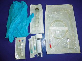
Although RVNs are not allowed to perform cystocentesis they should be aware of what is involved with the procedure including the equipment required (Figure 1). The equipment required for decompressive cystocentesis includes:
In most cases the presence of UO is a diagnosis in itself. Further abdominal imaging, including contrast radiography, may be indicated at a later stage.
Once the patient is considered stable, relieving the UO can commence. Depending on the patient’s status, sedation or general anaesthesia may be required.
Sacrococcygeal (SC) epidural blocks can be used to facilitate passage of a urinary catheter in cats with urethral blockage. A study of cats that presented with UO showed successful placement of the block (performed after opioid administration +/− sedative agent) may result in easier and more rapid unobstruction and placement of a urinary catheter (O’Hearn and Wright, 2011).
Additional observations include that cats appear in less pain post-catheter placement. The study concluded using an SC block can be a valuable adjunct to the management of feline UO in the emergency room setting.
To minimise complications (urethral rupture, uroabdomen and urethral strictures) associated with passing a urinary catheter, it is very important to extend the urethra caudally and dorsally before attempting urinary catheter placement.
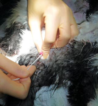
Prior to attempting to pass the urinary catheter, any mucous plugs may be able to be removed from the penile urethra by massaging the penis.
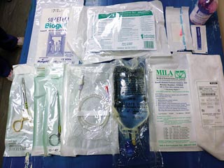
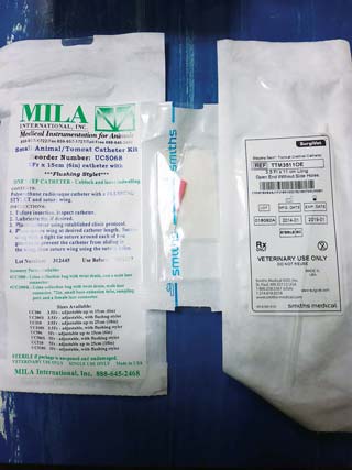
Equipment required for feline urinary catheter placement (Figure 2) includes:
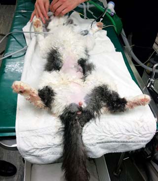
To “unblock” a cat, the sedated/anaesthetised patient should be placed in dorsal recumbency (Figure 4), then an area around the prepuce should be clipped and prepped. As previously mentioned, the urethral tip should be massaged to encourage dislodgement of any mucous plugs (any plugs should be kept for analysis).
A urinary catheter (preferably rigid and open ended) should be attached to a fluid extension set and 10ml syringe pre-filled with sterile saline. The system should be flushed to evacuate air and the catheter should be lubricated with sterile lubricant.
The urinary catheter tip should be introduced slowly into the urethral opening. As advocated by the SAFE approach, the urethra should be extended caudally (Figure 5) and the catheter then advanced slowly, until the tip lies in the bladder, while gently flushing with saline. Urine can then be removed from the bladder and the bladder can be lavaged with copious amounts of sterile fluid.
Once the urethra is cleared, a more suitable catheter to remain in-dwelling can be passed (softer and side holes).
There is controversy regarding catheter type and size – the more rigid catheters with end holes tend to help the effort to relieve the UO, but the rigidity can itself result in urethral trauma. If left in-situ they can cause more irritation compared to other materials so are not recommended as in-dwelling catheters. The softer catheters with side holes are preferred for in-dwelling use as they cause less irritation and are less likely to become blocked.
A review of controversies in the management of feline UO looked at catheter size. While it is recognised more research is needed, there is evidence from a small study to suggest a smaller (3.5Fr) urethral catheter may be associated with a decreased risk of reobstruction (Hetrick and Davidow, 2013).
Almost 50% of cats develop massive increases in their urine output following UO – a phenomenon called post-obstruction diuresis (POD; Balakrishnan and Drobatz, 2013). Increased rates of IVFT will be required to keep up with the patient’s urinary losses to prevent dehydration. The patient should be monitored for volume overload.
A closed system collection bag allows for accurate calculation of urine output (Figure 6), allowing the patients “ins and outs” to be monitored so IVFT can be tailored to suit the individual patient’s needs.
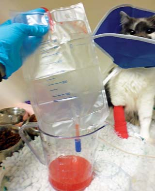
Commercial closed collection systems are available, but, alternatively, a system can be made with an empty fluid bag and fluid administration set. Patient interference with the urinary catheter should be avoided and the use of Elizabethan collars maybe indicated (Figure 7).
Another controversial area in the management of these cases is how long to leave the urinary catheter in-situ. Some argue the urinary catheter should be left in-situ for as short a time as possible, as the presence of a catheter can itself cause irritation and increase the risk of developing a urinary tract infection. Ideally, the duration of catheterisation should be determined by the clinical picture. This should factor in the presence of azotaemia, gross character of the urine and POD.
The prognosis for survival to discharge in most cats with UO is good, even in critically ill cats, providing they are stabilised within the first few hours of presentation (Balakrishnan and Drobatz, 2013).
There is no cure for FIC. There are reported recurrence rates of 15% to 40% in cases of feline UO (Cooper, 2015), so it is vital to implement measures to reduce the chance of recurrence. Environmental stressors have been reported to exacerbate clinical signs of FIC.

In cats with severe FIC, increased concentration of circulating catecholamines have been reported compared to control cats during a period of mild stress (Westropp et al, 2006).
Multimodal environmental modifications (MEMO) therapy can be very useful and the RVN can play a vital role in the success of these therapies. MEMO therapy begins with obtaining a thorough environmental history from the owners.
Questions to ask include (but are not limited to):
Once all the information is gathered, a tailor-made plan of preventive recommendations can be devised to meet the individual needs of the owner and cat. It is recommended to make only one or two changes at a time so as to not overwhelm the owner or cat. Advice may include the following.
The feline urethra has both smooth and striated muscle components, so drug combinations are often beneficial. Smooth muscle relaxants include acepromazine and phenoxybenzamine. Striated muscle relaxants include diazepam and dantrolene.
Nutraceuticals are generally aimed at protecting/repairing the glycosaminoglycan layer lining the bladder. It is thought this layer becomes damages with FIC.
It is thought pheromones induce changes in the limbic system and hypothalamus resulting in alterations in the emotional state of the animal. Cats have several pheromone-producing areas, including facial area, foot pads and perianal areas. A cat rubbing its face on its environment releases pheromones, which provides the cat with reassurance. Feliway (Ceva) is a chemical copy of one of the facial pheromones.
In conclusion, FLUTD is a common, but often complex, disease and the registered veterinary nurse can play a valuable role in the management of these cases, in both the acute and chronic stage.
Kerry Hall
Job Title