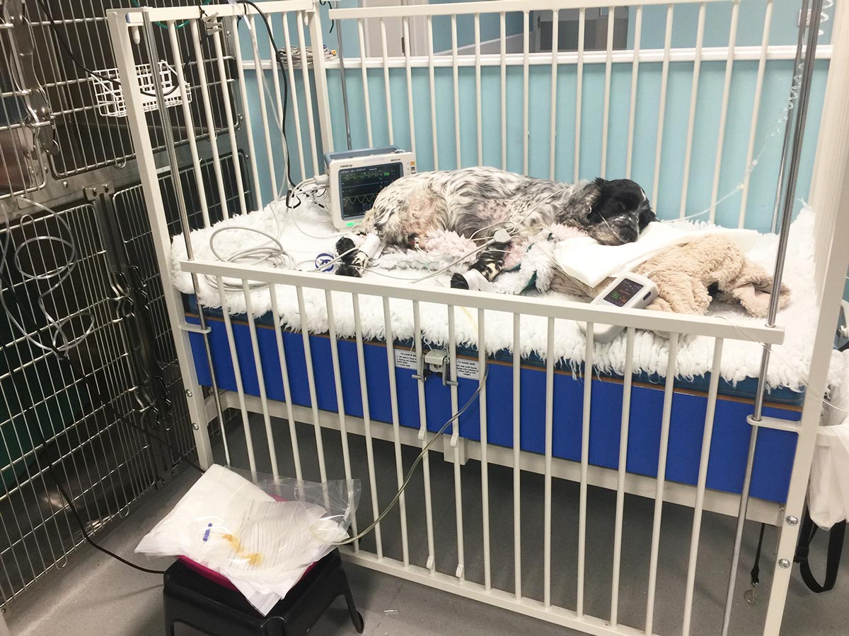
Urinary catheters in dogs – placement and maintenance tips
Louise O’Dwyer highlights the measures that need to be taken to prevent infection, both during and after catheterisation.
Louise O'Dwyer
Job Title
IMAGE: Sherry Young/Fotolia.
IMAGE: Sherry Young/Fotolia.
The placement of an indwelling urinary catheter is a commonly performed procedure in veterinary practice, and numerous different occasions and reasons why the procedure may be performed exist.
Despite it being a “common” procedure, the incidence of hospital-acquired urinary tract infections (UTIs) is common, so specific protocols should be established. These will ensure the procedure is performed aseptically and the device is maintained as aseptically as possible once in-situ. In turn, this should minimise the incidence of UTIs.
Indications for placement
It is appropriate to place a urinary catheter on a variety of occasions. These include:
Monitoring urine output – particularly in patients with renal disease, where hypotension or dehydration is a concern.
In situations where an inability to urinate, or urinary retention, may be a concern – for example, spinal disease and post-epidural administration (Welsh, 2003).
Drainage of the bladder prior to abdominal, vaginal and urethral surgery (Busch, 2006).
To achieve, or maintain, urethral patency in patients with urinary obstruction, or for hydropulsion in patients with urinary obstruction (Aldridge and O’Dwyer, 2012).
For the introduction of drugs or contrast agents for radiographic studies.
Urinary catheter types
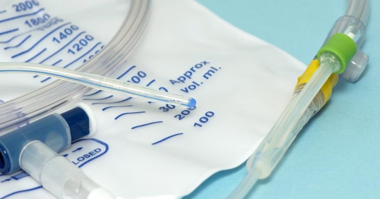
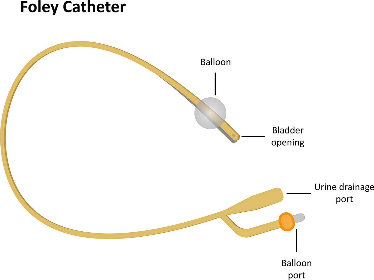
A wide variation in urinary catheter types, sizes and materials exists. Most practices are likely to stock Foley catheters (Figure 1), which are generally used as indwelling catheters.
Figure 1. A Foley catheter. IMAGE: joshya/Fotolia.
They feature a balloon at their distal end that becomes filled with saline once in situ to retain the placement of the catheter in the bladder. This reduces the need to suture the catheter to retain its position.
Polypropylene urinary catheters are also commonly used, but are most likely to be selected for intermittent catheterisation, rather than for indwelling catheters, therefore making them most suitable for:
obtaining urine sample collection
bladder emptying prior to surgical procedures
relieving urinary obstructions
These catheters are very stiff, therefore making them uncomfortable for the patient. This is why they are unsuitable to be placed as indwelling catheters.
Placement procedure
The procedure for placing a urinary catheter is as follows:
In lateral recumbency, hair is clipped from the vulvar area or preputial opening to maintain a hair-free area of at least 5cm away from the catheter insertion site.
The area is prepared using chlorhexidine scrub, which should be diluted to a concentration of 0.05%. Aseptic technique is ideally maintained by using sterile barrier drapes, surgical gloves and water-based lubricating jelly.
Female procedure
In females:
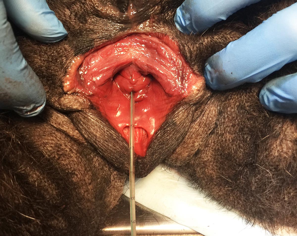
The vaginal vault is flushed with 2ml to 10ml of a 0.05% chlorhexidine solution for a minimum of five times. To insure appropriate placement, pre-measure the catheter along the path of the urethra to the bladder (Sullivan et al, 2010). Once aseptic preparation has been performed, sterile gloves should be worn (Davis and Riel, 2010).
A small amount of water-soluble lubricant should be placed on your finger and on the catheter tip. One obvious, but vital, consideration when catheterising female dogs is to ensure familiarity with anatomy. The valvular vault has three “levels”, with the urethral opening being found in the middle “vault” and the bottom “vault” being blind-ending (Thorpe, 2016).
Once happy with the location, the finger of the operator should be inserted into the vulva until the pelvic brim is palpated, and the urethral papilla and urethral opening sits just in front, or at the level of, the pelvic brim.
Once the urethral papilla has been located – on the midline of the ventral surface of the vulva – the tip of the finger should be curled just behind the structure. The catheter should then be fed along the underside of the finger towards the urethral opening, with the finger placed behind the papilla to guide the catheter downwards and into the urethra.
At the same time, the non-dominant hand should be used to advance the catheter into the vulva and then into the urethra. If the catheter is felt sliding past the finger, it should be withdrawn and placement into the urethra reattempted. Once correctly located, urine should be seen flowing from the catheter, or a syringe can be used to aspirate urine and confirm correct placement in the bladder (Figure 2). At this point, the balloon on the Foley catheter should be inflated with the correct volume of saline (the volume will be stated on the catheter itself or on the catheter packaging).
The catheter is often secured to the tail or a pelvic limb to minimise tension on the catheter itself. At this point, the catheter should be wiped down using a 0.05% chlorhexidine solution – an aspect of catheter care that should be regularly performed as part of maintenance.
Figure 2. Correct location of a urinary catheter in the urethra of a female dog.
Male procedure
In males:
After cleaning the extruded penis of any gross exudate, the prepuce is flushed with 2ml to 10ml of 0.05% chlorhexidine solution five times, ensuring you wear examination gloves.
Sterile gloves should then be worn while, with the penis still extruded, an appropriately sized urinary catheter is lubricated with water-soluble gel and advanced into the urethral opening into the bladder in an aseptic fashion. Urine flowing from the catheter confirms placement and further advancement of the urinary catheter can then be stopped. If the bladder is empty, flushing and aspirating sterile saline from the catheter can support proper placement, along with imaging (radiographs and/or ultrasound), if a question of proper placement still exists. A sterile closed collection system is immediately connected to the catheter following placement.
Management of indwelling urinary catheters
Indwelling urinary catheters should ideally be used as part of a “closed” system, which means the urinary catheter is not open to the outside.
Leaving catheters open to the air will increase the incidence of UTIs due to bacterial migration over the surface of the catheter. It can also result in urine scalding (Bubenik and Hosgood, 2008).
For maintenance, a urinary catheter can either be intermittently drained (via placement of a sterile bung at the end of the catheter with a syringe used to intermittently empty the bladder), or it can be connected to a sterile urinary collection bag.
The following procedure should be performed for the maintenance of indwelling urinary catheters attached to urinary collection bags:
Hands should be washed using the World Health Organization hand hygiene method and examination gloves worn throughout the procedure. Gloves should be changed and hand hygiene performed if contamination is considered to have occurred (Sullivan et al, 2010).
The insertion site of the catheter should be checked – if any signs of infection, discharge, inflammation, bruising or excessive discomfort are present, alert the clinician in charge of the case.
A gauze swab soaked in 0.05% chlorhexidine should be used to wipe the surface of the urinary catheter from the point of insertion to the collection bag.
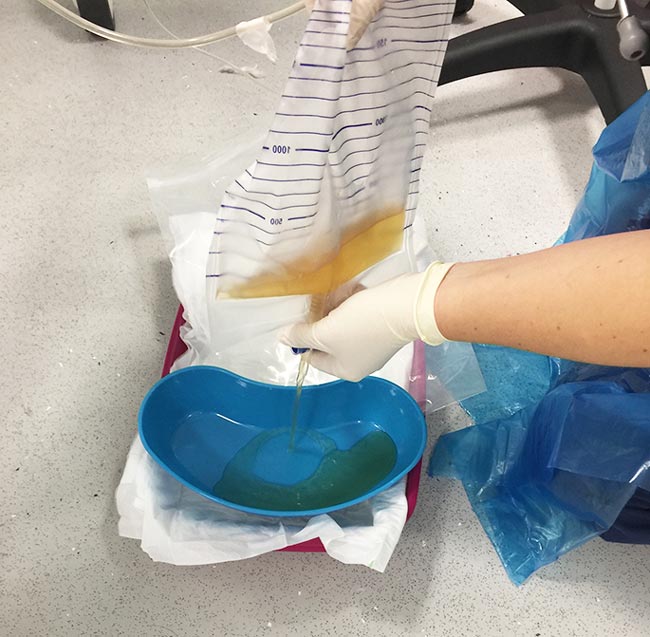
The bag may now be emptied into a urine collection jug. A surgical spirit-soaked swab should be used to wipe the bung before its removal to empty the urine collection bag, and again to wipe before and after replacement (Figure 3).
Figure 3. Correct personal protective equipment and technique used for emptying a urinary collection system.
If you have any concerns with the patency of a urinary catheter, it may be flushed with 5ml to 10ml of sterile water. This should only be performed if patency is queried to decrease the risk in infection due to regular disconnecting.
The vulva or prepuce should be cleaned and flushed with 0.05% chlorhexidine and, again, the urinary catheter should be cleaned from the insertion point to the collection bag once checks are complete – again, with chlorhexidine solution.
Once all checks are complete, the specific gravity, appearance and volume (including total volume and calculation of ml/kg/hour) of the urine should be noted on the patient’s hospital records. This information will give an indication of the patient’s renal function, infection and hydration status. Additional useful tests can include the performing of a urinary dipstick test.
The urine collection bag should ideally be placed inside a ziplock bag and/or rested in a clean tray lined with an incontinence sheet at a point lower than the patient (Figure 4; Smarick et al, 2004).
Figure 4. Placement of a urine collection system located within a ziplock bag and raised from ground level to minimise contamination.
Conclusion
Urinary catheterisation of dogs is a common procedure VNs can perform. In female dogs, this procedure is one often considered somewhat difficult to achieve, but with appropriate training and practical experience, it is achievable.
Whenever urinary catheters are being placed and maintained, an aseptic technique must be adhered to. Therefore, the use of a specific standard operating procedure is useful to act as a reminder to staff, and also to achieve consistency across the whole clinical team.
References
Aldridge P and O’Dwyer L (2012). Practical Emergency and Critical Care Veterinary Nursing, Wiley-Blackwell, Chichester.
Bubenik L and Hosgood G (2008). Urinary tract infection in dogs with thoracolumbar intervertebral disc herniation and urinary bladder dysfunction managed by manual expression, indwelling catheterization or intermittent catheterization, Vet Surg37(8): 791-800.
Busch SJ (2006). Small Animal Surgical Nursing: Skills and Concepts, Elsevier, Philadelphia: 38-39, 302.
Davis H and Riel D (2010). Diagnostic sampling and therapeutic techniques. In Bassert JM and McCurnin DE (eds), Clinical Textbook for Veterinary Technicians (7th edn), Saunders Elsevier, St Louis: 592-593.
Smarick SD, Haskins SC, Aldrich J et al (2004). Incidence of catheter-associated urinary tract infection among dogs in a small animal intensive care unit, J Am Vet Med Assoc224(12): 1,936-1,940.
Sullivan LA, Campbell VL and Onuma SC (2010). Evaluation of open versus closed urine collection systems and development of nosocomial bacteriuria in dogs, J Am Vet Med Assoc237(2): 187-190.
Thorpe A (2016). How to catheterise the female canine, Vet Nurse7(2): 121-123.
Welsh E (2003). Anaesthesia for Veterinary Nurses, Blackwell Publishing, Oxford: 215, 255, 261.