1 Sept 2021
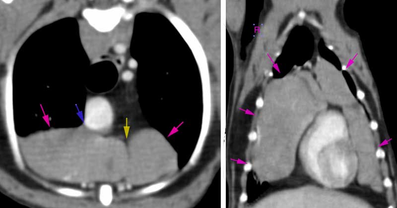
Tess was brought into ChesterGates Veterinary Specialists with coughing and lethargy, with CT scan revealing large mass occupying entire cranial mediastinum.
Joshua Silverwood
Job Title
A terrier that had a mass in its abdomen so large it was pushing against the heart and lungs has been saved thanks to vets at a Cheshire practice.
Tess the terrier was brought into ChesterGates Veterinary Specialists with coughing and lethargy, but a CT scan revealed a 6cm × 10cm × 8cm mass occupying her entire cranial mediastinum.
Vets diagnosed it as a thymoma and anaesthetised the dog to prepare for surgery. Referral surgeon Emma Hasleton, assisted by veterinary intern Anita Economou, performed a median sternotomy and tumour removal.
Dr Hasleton said: “This was a delicate and intricate process, because it was attached to her pericardium and cranial lung lobe.
“Tess’ owners were understandably absolutely devastated with the diagnosis. They did their research when we confirmed the cytological diagnosis after the fine needle aspirates, and they read a lot about these tumours and the risks of surgery.
“We talked at length about the significant risks associated with a thoracotomy procedure and the risk of Tess dying, and that once we could visualise the tumour, it may become apparent that it had invaded surrounding structures making it impossible to remove.”
Dr Hasleton added: “They were prepared for the possibility we would have to abandon surgery and not wake Tess up, if this was the case, so there was a real sense of uncertainty about the surgery for them. The whole team at ChesterGates was prepared on the day of surgery to focus solely on Tess.
“With these tumours you never really 100% know what you’ll find until you can visualise the tumour, and assess how it has grown and whether it has invaded or wrapped itself around any other structures in the chest, so there is always an element of uncertainty prior to surgery.
“In this case Tess was extremely lucky that there was no evidence of invasion – and although it was a huge tumour in this size of dog, it was able to be resected in full.”
Concluding, Dr Hasleton said: “Postoperative histology and immunohistochemical staining confirmed this was a lymphoid-rich thymoma, so her prognosis is excellent.
“Tess is doing really well post-operation. Her coughing is continually improving and she is back to her normal playful self, according to her owners.”