25 May 2015
Aimi Duff
Job Title
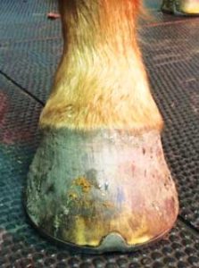
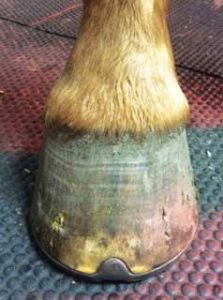
Figure 3. Long-toe, low-heel foot conformation associated with palmar foot pain.
Lameness is a common clinical presentation in horses. In the National Equine Health Survey 2014, lameness as a syndrome affected 18.5% of horses, with “foot lameness recorded in 4.6% of returns” (Slater, 2014).
External wounds or multi-limb lameness might complicate the case, but, by adopting a standard approach and with judicious use of diagnostics, the cause of lameness can be established. This paper details the approach to lameness pertaining to the equine foot.
As with any clinical problem, a full clinical history should be sought, with consideration for the following:
Clinical examination may reveal heat in the affected foot, which may be localised or diffuse across the hoof wall and coronary band (Milner, 2011; Munroe and Weese, 2011; Stephenson, 2011). There is, invariably, an increased digital pulse, with possible oedema extending proximal to the foot in the pastern, fetlock and cannon regions (Stephenson, 2011).
Detailed examination requires visual assessment of the foot in a resting state, carefully evaluating for visible abnormalities such as hoof wall cracks, avulsions and poor horn quality. Conformation and evidence of asymmetry with the contralateral foot should also be evaluated (Figure 1a and 1b). On closer inspection, hoof wall flare, medial-lateral imbalance (Figure 2) and circumferential event lines, or growth rings, might be seen (Munroe and Weese, 2011).
The degree of lameness is often variable depending on the aetiology. Hoof testers can be used to apply focal pressure to the sole, hoof wall, frog and heel regions; this can help in localising the painful area (Stephenson, 2011). Where a pain withdrawal response is elicited, any shoe might be removed to allow gentle paring, which may reveal sole defects or bruising (Munroe and Weese, 2011; Stephenson, 2011).
The affected limb should be carefully manipulated as pain or any restriction in range of motion might be associated with foot pain – for example, deep digital flexor tendinitis (Munroe and Weese, 2011).
In cases of mild lameness, gait analysis and evaluation of foot placement and break over can be useful for identifying factors that might contribute towards mechanical laminitis and collateral or sesamoidean ligament strains.
Perineural and intra-articular anaesthesia can help further localise the source of pain – for example, distal interphalangeal joint, palmar digital nerve and abaxial sesamoid nerve block (Milner, 2011; Munroe and Weese, 2011). It should be remembered local anaesthetic agents can diffuse from the site of injection, reducing the specificity of this technique for pain localisation (Nagy et al, 2009). Patients should therefore be evaluated within minutes of anaesthetic injection (Munroe and Weese, 2011; Nagy et al, 2009).
Diagnostic imaging may be necessary when physical examination does not yield a diagnosis. Radiography is useful for identifying fractures of the third phalanx (P3) or navicular bone, pedal osteitis, penetration wounds, interphalangeal collateral ligament injury, “rotation” of the third phalanx and so on (Munroe and Weese, 2011).
Projections used include dorsopalmar/plantar, lateromedial, 45º dorsoproximal-palmarodistal oblique of P3, 60º dorsoproximal-palmaro distal oblique and palmaroproximal-palmarodistal oblique of whole foot (Munroe and Weese, 2011).
MRI, scintigraphy and CT can be useful, although may be financially limiting. Scintigraphy can be used to confirm the significance of radiologic findings or detect early abnormalities that may not yet be visible radiographically. MRI is highly sensitive for detecting soft tissue pathology that would otherwise go undetected (Milner, 2011; Munroe and Weese, 2011).
The differential diagnoses for foot lameness include the following – some of which are detailed further on:
(Milner, 2011; Munroe and Weese, 2011).
Recurrent low-grade trauma due to conformational weakness or a forceful contusion can cause extravasation of blood from damaged vessels into the surrounding tissues and the formation of a “bruise”. This may be visible on the solar surface as an area of red discolouration. Bruising in the angle of the sole may be caused by poor or infrequent farriery. Solar micro fractures might enable secondary infection of the bruise and subsequent abscess formation (Munroe and Weese, 2011).

Lameness associated with bruising is variable. Management of bruising requires removal of the underlying cause – that is, providing padding over thin flat soles and addressing foot imbalance, excessive toe length and under-run heels (Figure 3). In acute bruising causing severe lameness, box rest and immersing the foot in cold water to reduce inflammation, along with concurrent NSAIDs, may be necessary (Munroe and Weese, 2011).
White line separation or solar defects might allow environmental bacteria to invade the underlying tissues, which can result in focal accumulations of purulent material (Milner, 2011; Munroe and Weese, 2011). Abscesses are often seen in horses with poor horn quality, or thin soles prone to bruising (Munroe and Weese, 2011).
The low compliance of the hoof capsule means the abscess exerts pressure on the sensitive structures within the foot, causing intense pain. This pressure can cause separation between the keratinised and germinal epithelial layers as the abscess tracks across the sole, or proximally to the coronary band where it may spontaneously burst (Milner, 2011; Munroe and Weese, 2011).
Lameness is often severe and affected horses may present with concurrent cellulitis in the affected limb (Milner, 2011; Munroe and Weese, 2011). Abscess tracks can be located using hoof testers and then gentle paring is used to reveal and open the track, enabling drainage (Munroe and Weese, 2011; Stephenson, 2011). It may be necessary to remove under-run sole to facilitate drainage (Milner, 2011; Munroe and Weese, 2011).
The mainstay of treatment is providing drainage and bandaging the foot until drainage ceases and lameness is resolved (Milner, 2011; Munroe and Weese, 2011). Drainage usually takes a few days, but if clinical signs fail to improve, radiography should be performed to check for deeper tissue involvement (Munroe and Weese, 2011; Stephenson, 2011).
Inflammation of the laminae within the foot is a clinical syndrome associated with a multitude of aetiological factors:
(Cripps and Eustace, 1999; Hood, 1999; Knottenbelt and Pascoe, 1994; Munroe and Weese, 2011).
The common denominator of these varied aetiologies is a disturbance in laminar blood flow; the exact pathophysiology remains a subject of intensive research, which is beyond the scope of this article (Hood, 1999; Knottenbelt et al, 1994).
Laminitis may be peracute, acute or chronic in nature. Acute laminitis manifests as severe pain in the front or all feet with bounding digital pulses, heat and reluctance to move. The horse may stand with a sawhorse stance and shift weight to offload the painful areas. The acute laminitic patient may be found recumbent (Hood, 1999; Knottenbelt et al, 1994; Munroe and Weese, 2011).
Movement is characterised by a heel first landing; a stilted, shuffling gait and pain – especially on turning (Hood, 1999; Knottenbelt et al, 1994). A pain withdrawal response is provoked when pressure is applied to the toe region using hoof testers. Altered weight loading, causing excessive deep digital flexor tendon tension along with dorsal laminar separation, can result in “rotation” of the pedal bone and, in very severe cases, “sinking” of the pedal bone within the hoof capsule and potential prolapse through the hoof sole (Hood, 1999; Knottenbelt et al, 1994; Munroe and Weese, 2011).
Chronic laminitis is characterised by pedal bone “rotation”, white line separation, dorsal hoof wall thickening and diverging rings (Hood, 1999; Knottenbelt et al, 1994; Munroe and Weese, 2011). Laminitis is managed by removing the underlying cause, controlling the associated neuropathic pain using NSAIDs and, in severe cases, opioids, gabapentin and lidocaine, remedial farriery and rest (Hood, 1999; Munroe and Weese, 2011).

Hoof cracks can occur in a horizontal or vertical plane as a consequence of foot imbalance, poor horn quality or trauma to the coronary band (Buffa et al, 1992; Josseck et al, 1995; Munroe and Weese, 2011; Figure 4). Cracks may be partial or full thickness and can be incomplete or complete depending on whether they extend to the coronary band (Munroe and Weese, 2011). Where they are associated with hoof wall instability, they can cause lameness. Cracks might also allow infection of the deeper tissues (Buffa et al, 1992; Josseck et al, 1995; Munroe and Weese, 2011).
Horizontal cracks may be seen as a consequence of abscesses that burst and drained at the coronary band, or in association with selenium toxicity (Milner, 2011; Munroe and Weese, 2011). Radiography may be necessary to diagnose secondary pathology of the third phalanx (Munroe and Weese, 2011). Treatment involves addressing any hoof imbalance and potential dietary biotin supplementation (Buffa et al, 1992; Josseck et al, 1995; Munroe and Weese, 2011).
In severe cases, debridement of the affected area is necessary to remove contaminated necrotic tissue before thoroughly cleaning the area and packing with resin or applying a plastic patch, plate or hoof staples to stabilise the area (Munroe and Weese, 2011; Pardoe and Wilson, 1999). A shoe with appropriately placed clips might also be used (Munroe and Weese, 2011).
Anaerobic bacterial infection of the frog can cause keratolysis of the frog, known as “thrush” (Munroe and Weese, 2011). This condition is associated with poor foot care; feet not picked daily, standing in unhygienic bedding and excessive moisture under remedial foot pads (Balch et al, 1997; Munroe and Weese, 2011). The frog and central sulcus become black with a characteristic malodour. The area between the heel bulbs can become affected, causing lameness (Munroe and Weese, 2011).

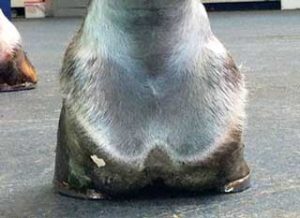
Management requires debridement of the affected tissue, improved foot hygiene and use of topical antiseptics such as iodine solution or hydrogen peroxide (Munroe and Weese, 2011). Remedial farriery may be needed if there is a conformational predisposition – for example, contracted heels (Figure 5).
Canker is a rare, painful, proliferative pododermatitis of uncertain aetiology, characterised by finger-like projections and a creamy exudate originating from the frog (Brandt et al, 2011; Munroe and Weese et al, 2011). Anaerobic bacterial and bovine papilloma virus infection are thought to be causal (Munroe and Weese, 2011). Lameness is variable depending on the severity of disease.
Management requires debridement of the affected tissue followed by dressing with wet-to-dry antibiotic dressing using metronidazole and possible concurrent systemic antibiosis (Brandt et al, 2011; Munroe and Weese, 2011).
Epithelial tumours of the hoof germinal layers are known as keratomas and the exact aetiology for them is uncertain. As the tumour increases in size, it causes hoof capsular distortion and pressure lysis of the solar margin of the third phalanx with associated lameness (Boys Smith et al, 2006; Munroe and Weese, 2011).
Dorsoproximal-palmarodistal radiography projections support the diagnosis, which can be confirmed on histological examination of biopsy samples.
Surgical excision following resection of the overlying hoof wall carries a good prognosis for recovery, although tumour recurrence, hoof wall instability and the formation of exuberant granulation tissue are possible complications (Boys Smith et al, 2006; Munroe and Weese, 2011).
Bacterial and fungal infection can cause keratolysis of the stratum medium of the hoof forming cavities. If extensive areas of hoof wall are involved, support for the distal phalanx is lost and it may displace, causing lameness and irreversible phalangeal changes (Munroe and Weese, 2011; O’Grady, 2002; Turner, 1998).
Radiography is indicated to confirm the extent of white line involvement and detect phalangeal changes, prior to debriding all affected tissue. The affected area should then be cleaned thoroughly using 2% iodine solution and shod appropriately to provide pedal support (Munroe and Weese, 2011; O’Grady, 2002; Turner, 1998).
The third phalanx may fracture following trauma, with the fracture plane following several different possible configurations involving the body and/or wings. These fractures are further classified as articular or non-articular, depending on whether they involve the distal interphalangeal joint. Patients with phalangeal fractures present with acute onset lameness of variable severity, depending on the configuration (Honnas et al, 1988; Munroe and Weese, 2011).
Lameness may become chronic in nature with articular fractures. Diagnosis is confirmed with diagnostic imaging such as radiography, CT, scintigraphy or MRI (Honnas et al, 1988; Munroe and Weese, 2011).
Management requires strict rest and minimising hoof capsular movement to facilitate healing. This can be achieved by fitting a bar shoe and solar packing, (Honnas et al, 1988; Munroe et al, 2011).
Intra-articular steroids may be needed to help manage secondary distal interphalangeal osteoarthritis (Munroe
and Weese, 2011).
Pedal osteitis is characterised by demineralisation of the distal phalangeal solar margin. This may occur secondary to chronic trauma, laminitis and pedal rotation, keratoma formation and chronic toe bruising. Pedal osteitis may be an incidental radiological finding secondary to historic disease or reflective of active pathology.
A detailed clinical examination including the application of hoof testers and possible perineural anaesthesia are needed to establish the significance of radiographic findings. Concurrent sub-solar abscessation may be associated with the development of septic pedal osteitis and possible sequestrum formation (Gaughan et al, 1989; Milner, 2011). Treatment for pedal osteitis requires that the inciting cause is removed, and where there is pedal sepsis surgical curettage may be necessary (Gaughan et al, 1989). Remedial farriery may be needed to provide support.