3 Oct 2016
Wayne McIlwraith discusses arthroscopic surgery in horses and highlights its principal advances in the past 10 years.
Wayne McIlwraith
Job Title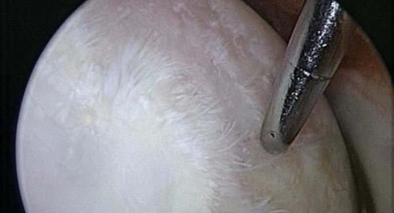
Figure 1a. Diffuse articular cartilage fibrillation and erosion on the medial femoral condyle of the stifle. Image: McIlwraith CW et al (2014)2.
Arthroscopic surgery has revolutionised orthopaedic surgery in the horse and our ability to return equine athletes to their full potential.
The first edition of Diagnostic and Surgical Arthroscopy in the Horse was published in 19841, detailing general technique and diagnostic arthroscopy, as well as diagnostic and surgical arthroscopy in the carpal, metacarpo/tarsophalangeal, femoropatellar and tibiotarsal (tarsocrural) joints. It was 135 pages.
The fourth edition, published in 20142, meanwhile, is 454 pages long, with 17 chapters. Within its pages, techniques in all the joints of the appendicular skeleton, as well as tendon sheaths and bursae, are described.

MRI has become an accepted and validated diagnostic technique in equine orthopaedics, with more specific diagnoses of conditions involving soft tissue leading to further indications for diagnostic and surgical arthroscopy.
A good example of this is lesions of the deep digital flexor (DDF) tendon associated with the navicular bursa and involving the dorsal surface of this tendon.
Lesions are identified by MRI, then approached endoscopically through the DDF tendon sheath into the bursa, as described in a study by Smith and Wright in 20123.
However, limitations have been recognised in both human and equine OA regarding the usefulness of MRI, with diagnostic arthroscopy remaining the gold standard for:
In the stifle, for example, the usefulness of MRI is very limited because the only resonance that allows for it to be contained and imaged is of low field strength.

In the femorotibial joint, meanwhile, arthroscopy remains the gold standard for diagnosis of soft tissue lesions, as well as definition of articular cartilage loss. Figure 1 shows examples of articular cartilage fibrillation with an area of erosion down to the calcified cartilage layer on the medial femoral condyle in the femorotibial joint (Figure 1a) and full thickness erosion with bone eburnation on the axial side of a medial femoral condyle in another femorotibial joint (Figure 1b) – arthroscopy was the only way of diagnosing these.
Similarly, while meniscal tears can commonly be diagnosed with expert diagnostic ultrasound examination, the definitive diagnosis of these tears and their treatment requires arthroscopic surgery (Figure 2) and commonly results in the horse returning to full athletic activity. The overlap between what areas of the femorotibial joint can be examined – comparing diagnostic arthroscopy and ultrasonography, in a study led by Myra Barrett4 at the Orthopaedic Research Center at Colorado State University – has also been defined.
Separate approaches to different compartments requiring careful technique has resulted in improved diagnostic ability. For example, in the femorotibial joint, examination in the cranial pouch of the medial femorotibial joint only allows for the cranial horn of the meniscus to be visualised. However, a specific approach to the caudal pouch in the medial femorotibial joint has enhanced our ability to diagnose lesions in the caudal horn and make inferences regarding lack of structural stability in the meniscus.

As mentioned previously, the conjunctive use of arthroscopy and ultrasonography provides additional information.
Our ability to return horses with carpal slab fractures and displaced distal metacarpal condylar fractures to full athletic activity has greatly improved, meanwhile, due to the careful combined use of a: needle placement under arthroscopic visualisation to define limits of intra-articular fractures, and b: radiographs for correct position and direction of lag screw placement. The ability to manipulate under arthroscopic visualisation also exists.
With slab fractures, the combination of a: arthroscopic evaluation and placement of needles – to define lateral to medial location for lag screw fixation – and b: radiographs to give exact positioning in the bone relative to the fracture and direction of the screw, allows for definition of the best position to place the screw in the dorsal surface, as well as direction and angle.
In the case of fractures of the metacarpal and metatarsal condyles, there is a move away from feeling the fracture has to be directly visualised through arthrotomy to get optimal reduction. By being able to arthroscope both the dorsal pouch and the palmar/plantar pouch, its exact congruency can be obtained with manipulation prior to clamping the fracture and then placing the lag screws (Figure 3).

There has also been considerable further documentation and validation of arthroscopic techniques for removal of osteochondral fragmentation, debridement of defects, as well as treatment of osteochondritis dissecans (OCD) lesions in the carpus, fetlock, femoropatellar, tarsocrural, and proximal and distal interphalangeal joints, allowing us to give more accurate prognoses in specific conditions in the joint. While much traumatic osteochondral fragmentation, as well as OCD lesions, are treated with removal and debridement, techniques have also been developed to lag screw larger osteochondral fragments in place. A technique has been developed to fix larger OCD flaps with absorbable pins (Figure 4), with very successful results.
Another condition where debate continues about the most effective treatment is subchondral cystic lesions (SCLs) of the medial femoral condyle in the medial femorotibial joint. The initial arthroscopic technique of debridement under arthroscopic evaluation has evolved into various options, including the intralesional injection of triamcinolone acetonide under arthroscopic guidance, which preserves the surface articular cartilage.
This technique was developed from basic research where the linings of SCLs were shown to have increased levels of deleterious mediators, resulting in osteoclastic resorption. It has been successful, but at the 90% level with cysts without any secondary OA when the lesion is unilateral, and at the 70% in bilateral cases for bilateral lesions. The prognosis is also decreased if spurs on preoperative radiographs are present.

Opinions diverge on the treatment of SCLs not responding to intralesional injection. Options include debridement, followed by use of cancellous bone grafting or bone substitute and the addition of stem cells in fibrin and/or platelet-rich plasma (PRP).
In recent cases, the author has adopted the technique described in a study by Santschi et al5 of lag screw fixation when intralesional injection has failed to provide athletic soundness – this has been successful in the few cases he has done.
Finally, a technique for standing arthroscopic evaluation of the equine stifle has been developed by Dave Frisbie. This was to obtain diagnoses in a group of athletic horses that had clinical evidence of stifle disease based on diagnostic analgesia and equivocal changes on radiographic and ultrasonographic examinations. However, owners were reluctant to elect for conventional arthroscopy under general anaesthesia. Therefore, the use of an 18-gauge (1.3mm) needle arthroscope in the standing sedated horse allows shortened post-procedure rehabilitation and can help identify presence or absence of a significant lesion. It is not a treatment modality and is described by Dr Frisbie in McIlwraith et al2.

An important part of achieving success with certain arthroscopy procedures is the adjunctive use of therapies to deal with secondary OA and articular cartilage lesions. The main advances in this area have been development and validation of biologic and stem cell therapies, and the state of these has been reviewed in the second edition of Joint Disease in the Horse6.
Biologic therapies include the protein-based therapies of autologous conditioned serum (ACS) and PRP. Evidence – in both the equine model of OA, as well as clinical cases – is positive for the value of ACS as an adjunctive treatment when there is osteoarthritic change present in the joint(s).
Documented evidence of the use of PRP in treating joint disease, meanwhile, has not been attained as yet, but some veterinarians are using it intra-articularly in OA. There is precedent here in that a number of publications have reported successful results with the use of intra-articular PRP in OA of the human knee.

While numerous recognised sources for mesenchymal stem cells (MSCs) exist, the most positive evidence has been developed with bone marrow-derived autologous MSCs. Following a landmark paper showing stem cell therapy in a caprine model of OA could facilitate regrowth of the meniscus, as well as decrease OA changes, we embarked on a multi-centre study following arthroscopy of femorotibial joints and showed enhancement of success rate, compared to arthroscopic surgery alone for both meniscal injuries as well as articular cartilage erosion (Figure 1).
Advances have also been made with physical rehabilitation techniques. Good scientific evidence has demonstrated the value of underwater treadmilling in decreasing clinical symptoms of OA, as well as improving proprioception, which we believe to be a critical factor in returning athletes back while minimising re-injury. All of these advances in physical rehabilitation techniques have also been reviewed3.