26 Mar 2024
In the latest of his Diagnostic Dilemmas series, Francesco Cian reviews the case of a 14-year-old Arabian gelding presented with a subcutaneous mass in the dorsal neck area.
Francesco Cian
Job Title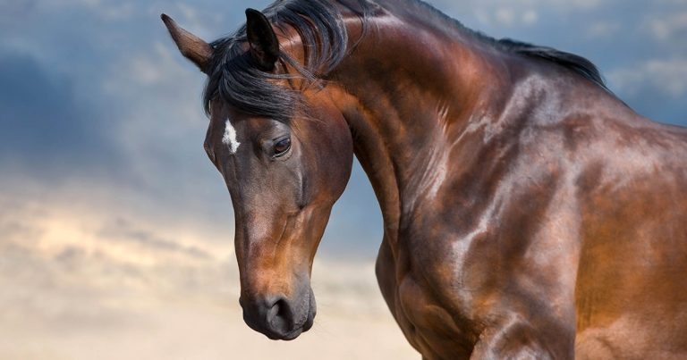
Image © kwadrat70 / Adobe Stock
A 14-year-old Arabian gelding presented to the referring vet for investigation of a firm, SC mass in the dorsal neck area that appeared three weeks prior. No other clinical signs were noted.
Cytological samples of the mass were obtained by fine-needle aspiration and were submitted to an external laboratory for examination.
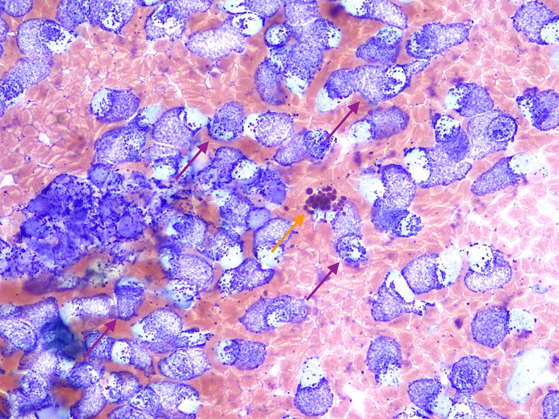
The sample (Figure 1) has high cellularity and adequate preservation. The background is clear with large numbers of red blood cells. A main population of discrete nucleated cells are present, characterised by numerous purple/magenta granules filling most of the cytoplasm and often obscuring the nuclei. These cells are referable to mast cells (purple arrows) and show mild anisocytosis (cell size variation).
Rare eosinophils (orange arrow) are also noted. These cells have large orange/brick red granules giving the cells in this species the typical raspberry-like appearance.
These cytological features were supportive of a mast cell tumour diagnosis. Surgical excision was performed and the sample was submitted for histopathological examination, which confirmed the initial cytological diagnosis.
At a six-month follow-up, the lesion had not recurred.
Mast cell tumours are relatively uncommon neoplasms in the equine species, contrary to what are observed in dogs, and represent 2% to 10% of all cutaneous tumours. They have been rarely described in other locations, such as the eye and the respiratory system.
A clear breed, age and sex predilection does not exist, but male adult Arabian horses may be over-represented. Lesions appear as distinct nodules of variable sizes – most often single – with intact overlying skin and occasional alopecia. Most common anatomic locations include the head, trunk and limbs.
These tumours typically demonstrate a slowly progressive or static growth pattern and prognosis is excellent with surgical excision. Rare cases of spontaneous regression have been documented. Given their usual benign behaviour, they have been also referred in the past with the term “mastocytosis”, originating from human medicine to define benign mast cell disorders.
Rare cases of local tissue invasion and metastases have been described, including very rare forms of multicentric forms.
Similarly to that observed in other species, the diagnosis of a mast cell tumour can be easily achieved with cytology by fine needle aspiration.
Did you know?
According to the latest National Equine Health Survey from 2018, the most frequent general disease syndrome recorded was skin disease, accounting for one third of all problems reported, followed by lameness (including laminitis) and metabolic disorders. Among the tumours, sarcoid and melanoma were the
most commonly reported.
Aspirates from these tumours are characterised by a main population of mast cells with variable degree of granulation and atypia. These may be accompanied by eosinophils, fibroblasts and/or collagen fibres, since the substances contained within mast cell granules may induce fibroplasia, collagenolysis and eosinophil chemotaxis.
Chronic lesions with significant eosinophilic inflammation may be difficult to differentiate from collagenolytic granuloma and parasitic eosinophilic granulomatous dermatitis.
Mast cell tumours are uncommon cutaneous tumours in horses.
Different from what is observed in dogs, the vast majority of equine mast cell tumours are invariably benign and can be cured with surgery alone. Metastatic disease is a very rare event.