5 Feb 2020
“Our ability to image the entire neck in such detail has significantly improved our understanding of neck pathology and, in some cases, has led to the development of new treatments and techniques” – RCVS specialist in equine surgery Russell Parker.
David Woodmansey
Job Title
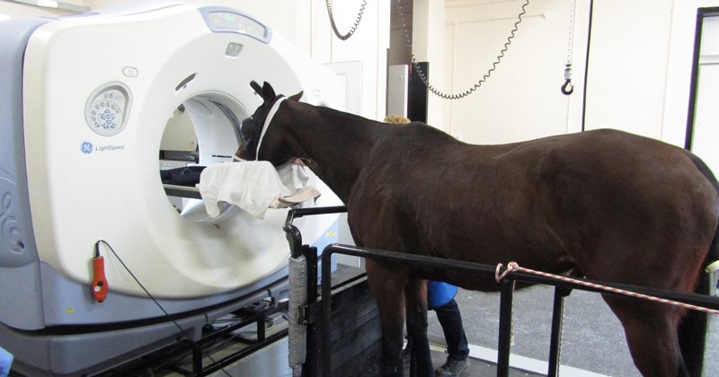
A horse undergoing CT scanning of the head. Image © Liphook Equine Hospital
Staff at the CT facility at Liphook Equine Hospital in Hampshire are celebrating after scanning their 500th patient.
The custom-built advanced imaging suite has its own padded recovery room and an adjustable hovering platform to accommodate the patient, and boasts an 80cm-wide CT gantry, allowing imaging of more of the animal than was previously achievable.
The majority of cases undergo scanning of the head and upper neck under sedation while stood on the platform, which can be adjusted to allow horses of all different sizes to be scanned – from Shetland ponies to shire horses.
In addition, horses can also be scanned under general anaesthesia, allowing imaging of the neck or limb in large horses, or the entire horse itself in the case of foals or smaller breeds.
Russell Parker, one of six specialist surgeons at Liphook, said: “Even for relatively routine cases of sinus or dental disease, this technology has improved our ability to make an accurate diagnosis and formulate an effective treatment plan.”
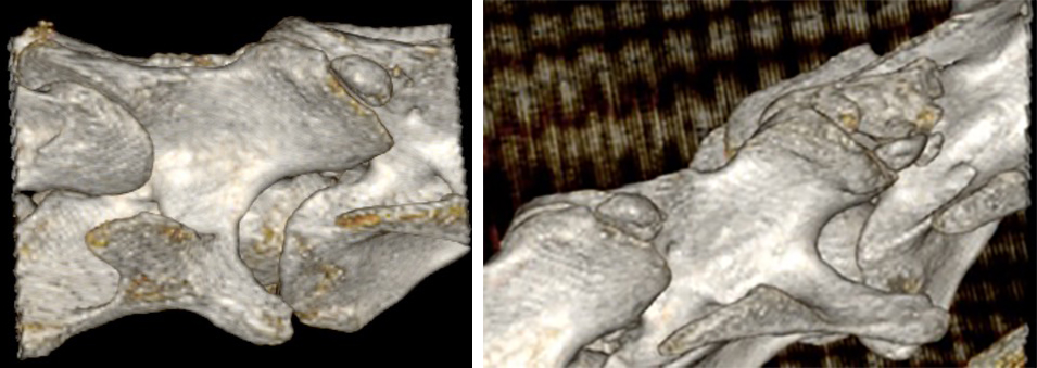
Since the facility opened in 2016, the team has also scanned more than 50 horses with neck-related issues caused by a wide range of conditions, such as cervical vertebral stenotic myelopathy (wobbler syndrome), fractures of the vertebrae and facet joint pathology.
Mr Parker continued: “Our ability to image the entire neck in such detail has significantly improved our understanding of neck pathology and, in some cases, has led to the development of new treatments and techniques, which is exciting for us as a surgical team.
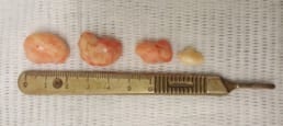
“For example, fragments within the cervical facet joints have barely been recognised up until now, but we have found them in approximately a quarter of the horses we have scanned and have now operated on four horses to remove them – all of which are doing well.”