15 Aug 2016
Moses Brennan and Rachel Agass discuss advances in a variety of techniques developed for treatment and closure of wounds present in horses.

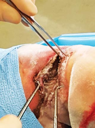
Figure 4. Appearance of the same wound in Figure 3 during debridement with a hydrosurgical wound debridement system. The wound was sutured post-debridement and placed in a fiberglass cast, resulting in good primary intention healing.
Equine wounds pose a number of therapeutic challenges. Primary closure of extremity wounds is seldom possible in equine patients due to the often marked area of tissue loss, excessive contamination, high levels of tissue tension and prolonged duration prior to presentation.
In fact, a retrospective study of 422 horses revealed primary closure being the treatment of choice in 24% of cases (Wilmink et al, 2002).
Equine distal limb wounds, in particular, exhibit ongoing inflammation, greater wound edge retraction and slower epithelialisation rates, as well as premature cessation of contraction and formation of exuberant granulation tissue, or “proud flesh” (Jacobs et al, 1984; Bertone, 1989). Many of these problems can be attributed to relatively poor vascular supply, the absence of muscle cover and the numerous bony prominences, as well as high levels of motion and often significant wound contamination (Schwarts et al, 2002; Cochrane et al, 2003).
The basic principles of wound management have been outlined elsewhere; this article will focus on advances in wound management.
Manuka honey has been used as a topical treatment for open wounds for thousands of years (Majno, 1975). Honey as a topical therapy in wound healing lost favour with the development of modern antimicrobial drugs; however, the emergence of bacterial resistance has prompted renewed interest in its use.
The Leptospermum species of plants includes about 83 species found mainly in Australasia. Honey derived from these plants has been shown to have superior antimicrobial activity compared to many other varieties (Snow and Manley-Harris, 2004). The Manuka bush (Leptospermum scoparium), found in New Zealand, has been studied most extensively and is used for the commercial production of medical grade manuka honey, with a standardised antimicrobial activity.
Methylglyoxal (MGO) is produced from dihydroxyacetone, which is found in high concentrations in the flower of the manuka bush; the MGO is responsible for the antimicrobial activity (Mavric et al, 2008).
Commercial batches of manuka honey are graded with a unique manuka factor (UMF). This rating compares the antimicrobial activity of the honey compared to an antiseptic versus Staphylococcus aureus:
Several studies have been conducted looking into the effects of the use of manuka honey of the process of second intention wound healing in the equine distal limb (Dart et al, 2015). Experimental wounds were created on the distal limb of standardbred horses and wounds were bandaged either with manuka honey or left untreated for 12 days.
The wounds treated with UMF 20 honey retracted less and remained smaller than untreated wounds until day 42; however, no difference occurred with overall healing time. These studies suggested a beneficial effect on second intention healing and the principal therapeutic effect appeared to be in the early stages of healing.
The importance of wound debridement and decontamination cannot be overemphasised. Early wound debridement is essential for removal of foreign material and reduction of bacterial load – both of which can be associated with inadequate or delayed wound healing (Hendrickson and Virgin, 2005). Foreign material reduces the bacterial numbers required to result in infection; thus, adequate decontamination and maximal preservation of viable tissue are of paramount importance.

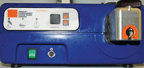
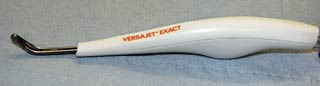
Debridement methods include sharp, mechanical, chemical and autolytic; conventionally, sharp and mechanical methods have been most widely used. Mechanical debridement methods usually consist of wound lavage with sterile saline at a pressure of 10lbs to 15lbs per square inch (PSI; Stashak, 2008). A method of mechanical hydrosurgical debridement has been developed (Figures 1 and 2). The unit works by generating a pressurised, high-velocity stream of sterile saline, creating a localised Venturi (suction) effect, facilitating removal of contaminated and necrotic tissue, while irrigating the wound (Stashak, 2008); therefore, selectively removing non-viable tissues.
This system has been shown to reduce in vitro S aureus load from contaminated equine muscle more successfully than conventional means (Skärlena et al, 2015) and is also frequently used in human medicine for debridement of burns and chronic wounds (Hyland et al, 2015; Liu et al, 2015). Figures 3 and 4 demonstrate the appearance of a heavily contaminated wound before and after hydrosurgical debridement.
Vacuum-assisted wound closure (VAC) – also known under the pseudonyms negative pressure therapy and the vacuum-sealing technique – is the application of sub-atmospheric pressure to a wound through an open pore foam dressing to assist healing (Lambert et al, 2005; Figures 5 and 6).

The VAC system consists of a polyurethane ether foam sponge (pore sizes ranging from 400μm to 600μm) cut to fit directly over the wound surface. An adhesive dressing is then placed over the sponge to fix it in place and trimmed so it overlaps the wound edge by 4cm to 5cm.
A small opening is created in the adhesive dressing over the sponge, over which a non-collapsible evacuation tube with a fenestrated distal end surrounded by an adhesive dressing is then placed. The fenestrations at the end of the tube establish communication between the lumen of the tube and the foam sponge.
The free end of the tube is connected to an adjustable vacuum pump, creating suction that allows subatmospheric pressure to be applied to the entire wound surface. The pressure can be applied in a constant or intermittent manner up to 125mmHg (Venturi et al, 2005).
Exposure of the wound bed to subatmospheric pressure results in increased local blood flow; a fourfold increase in local flow is observed when a pressure of -125mmHg is applied (Morykwas et al, 1997). This increase in blood flow is most marked in the first five minutes and the same increase is observed when negative pressure is reapplied after a short break, hence a regimen of five minutes on, two minutes off, was developed in human medicine.
In veterinary medicine, however, a continuous application of subatmospheric pressure is usually used as the initiation of the vacuum is deemed painful.

Negative pressure also stimulates cellular proliferation and increased mitotic activity in response to the mechanical stress exerted by the VAC system (Venturi et al, 2005). Wound exudate containing inhibitory proteases and collagenases are also removed from the wound surface (Goldsworthy and McAlinden, 2011). The net result of these mechanisms is accelerated granulation tissue formation.
VAC application significantly decreases bacterial load. Experimentally infected pig wounds (inoculate with 108 bacterial microorganisms) treated with VAC had a decrease in bacterial count by a factor of 103 over four to five days, with effectiveness being seen following inoculation with S aureus and Streptococcus epidermidis. This change was dramatically different from that seen in the control group, which had peak levels of bacteria approaching 109 organisms per gram on day 5, dropping to less than 105 after a mean of 11 days (Morykwas et al, 1997).
The ability of VAC to decrease bacterial load is thought to be due to improved blood supply, decreased interstitial oedema and removal of harmful enzymes from the wound bed (Venturi et al, 2005). Additionally, improved local oxygenation improves wound defenses and increases resistance to anaerobic infections.
The VAC system is widely used in human medicine and surgery in all but ischaemic wounds (Armstrong et al, 2004), having first been employed for the treatment of open fractures in the 1990s (Fleishman et al, 1993). The system has been used in veterinary medicine for some time, although the first reports for its use in canine traumatic wounds were seen in 2007 (Ben-Amotz et al, 2007), focusing on management of distal extremity wounds.
Commercial units are available and, although home-made systems can be made, they require a higher level of maintenance to ensure efficacy. A word of caution – theatre suction pumps are not designed to be used for this purpose and will easily overheat when used continuously (Goldsworthy and McAlinden, 2011).
The list of veterinary indications is ever-growing, including degloving/shearing wounds of the distal, avulsion wounds, chronic non-healing wounds, thoracic wall wounds/sternotomy wound dehiscence, assistance of wound contraction, securing skin grafts, treatment of cellulitis and seroma prevention (Goldsworthy and McAlinden, 2011).
Many of these indications are more commonly encountered in small animal practice; however, use in equine practice is growing. The first case report in an equine patient was in 2005, where the system was successfully used in the management of a traumatic neck wound (Gemeinhardt and Molnar, 2005).
The device can be applied over any type of tissue or material, including dermis, fat, fascia, tendon, muscle, blood vessels and bone, to name a few (DeFranzo et al, 2001). The wound must first be thoroughly debrided and free of necrotic tissue to avoid the risk of promoting a deeper or systemic infection (Gwan-Nulla and Casal, 2001); as such, the system is not usually applied until it is at least 24 to 48 hours old. The wound must also be well vascularised because further necrosis is likely to occur at the edges (Clare et al, 2002).
The sponge should be changed every 48 to 72 hours and this can sometimes be difficult because of the rapid in-growth of granulation tissue, necessitating more frequent changes.
Complications are rare, but can include desiccation of tissue if the seal is improper, dermatitis of surrounding skin, excessive in-growth of granulation tissue into the foam and loss of vacuum through dressing damage. VAC should not be applied to wounds where there are overt signs of infection, significant necrotic tissue, active hemorrhage, or if a neoplastic aeitiology is suspected or confirmed.

The ability of biological tissues to stretch and expand when placed under continuous stress for a prolonged period has been recognised in numerous physiological and pathological situations – for example, abdominal wall laxity during pregnancy.
First used in human medicine in 1957, the concept was developed with a rubber balloon used to expand the skin for reconstruction around an ear wound (Neumann, 1957). The technique re-emerged 20 years later and has been used in numerous plastic and reconstructive procedures in the human field. Conventional tissue expanders required manual filling through an external port in the subcutaneous tissue.
This process is painful for the patient and needs to be carried out by a trained professional. This lead to the development of self-inflating tissue expanders, consisting of hydrogel, vinyl pyrrolidone and methyl methacrylate that absorb the surrounding tissue fluid, and increase in size over a six to eight-week period (Weise, 1993).
Use of self-inflating tissue expanders is being trialled in equine patients. Although, as yet, no clinical research exists, except for an experimental study using a rabbit model (Timmenga et al, 1991). Several cases have been subjected to this management with success, with the treatment seeming most useful for reconstruction of distal extremity and head wounds of horses and small animals.

Soft tissue expanders are commercially available to vets. The authors have used the expanders with success in the removal of a supraorbital melanoma and subsequent closure of the surrounding skin, although a variety of other applications are possible (Figures 7, 8 and 9). Potential complications are few in number and frequency, but include pain and necrosis of the skin flap or underlying tissue.
The role of mesenchymal stem/stromal cells (MSCs) in wound repair has been relatively well characterised. MSCs are multipotent cells with the capacity to differentiate into various tissue-forming cell lineages, including chondrocytes, tenocytes, adipocytes among others (Krampera et al, 2006).
Continued research, in both human and veterinary fields, have lead to the clarification of the mechanism through which MSCs contribute to improved wound healing.
Classically, the primary beneficial effect in the regenerative process is thought to be the cellular differentiation restoring damaged tissue (Bussche et al, 2015).
More recent data has indicated paracrine signaling may be the primary mechanism for the regenerative effects observed through all three phases of wound healing (Baglio et al, 2012; Madrigal et al, 2014).
Paracrine signaling involves the release of biologically active molecules, which have effects on the survival, migration and proliferation of surrounding cells (Maxson et al, 2012). Their presence, therefore, supports healthy physiologic function.
Andiogenic signaling factors have been identified in equine MSC-derived conditioned medium (CM) with these factors capable of inducing cell proliferation and vessel formation in vitro (Bussche et al, 2014).

CM derived from MSCs contains factors secreted by the cells, without the cells themselves. This implies CM obtained from cell cultures may be used as a “stem cell free” therapy in regenerative medicine; therefore, avoiding the potential negative effects that may arise with the use of the cells themselves, including inherent toxicity, risk of tumour formation and allograft immune rejection (Haarer et al, 2015; Maguire, 2013).
A study conducted by Bussche et al (2015) investigate the in vitro effect of equine MSC-derived CM on cellular biology. The MSCs were harvested from peripheral blood and the resulting CM was delivered to tissue in a hydrogel microcapsule.
The authors discovered the CM stimulated dermal fibroblast migration and positively influenced the expression of those genes involved in wound healing; therefore, theoretically improving equine cutaneous wound healing.
Interestingly, a suggestion was also made that the effect achieved was donor specific, suggesting donors may be screened and selected based on the desired therapeutic outcome. Clearly, this research is very much in its infancy; however, it does provide an interesting possibility for the future.