7 Mar 2019
Finding a cause of equine lameness can be straightforward, but often evaluating poor performance and reaching a consensus on it can be a complex and time-consuming process for first-opinion practitioners.
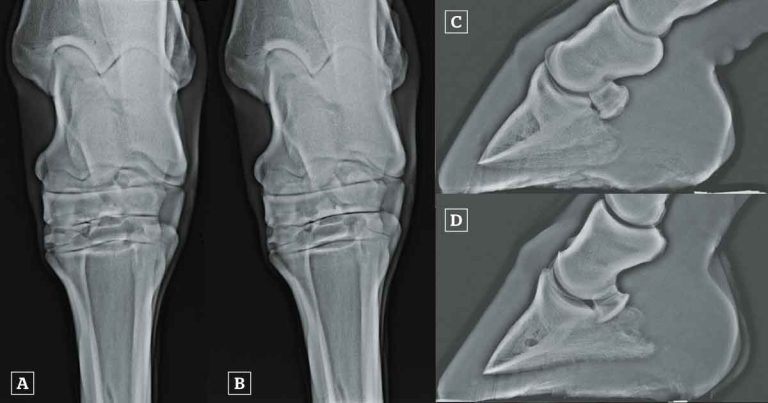
Figure 3. [A] Dorsoplantar radiograph of a hock acquired with a horizontal beam. The centrodistal and tarsometatarsal joint spaces are poorly defined (arrows). Artefactual narrowing of the distal tarsal joints is common in dorsoplantar images acquired with a horizontal beam because the distal tarsal joints slope from proximal, dorsally to distal, plantarly. [B] Dorsoplantar radiograph of the same hock as in A, acquired with a beam angled 5° distally (dorsal 5° proximal-plantarodistal oblique). The centrodistal and tarsometatarsal joint spaces are clearly visible and no narrowing exists. [C] Lateromedial radiograph of a foot of non-diagnostic quality. This is not a true lateromedial because the distal condyles of the middle phalanx and the palmar processes of the distal phalanx are not superimposed. The demarcation between the palmar compact bone of the navicular bone and the spongiosa, and the integrity of the palmar compact bone, cannot be assessed, because the beam is not perpendicular to the long axis of the navicular bone. The navicular bone cannot be evaluated using this image. [D] Lateromedial radiograph of the same foot as in C, acquired with better positioning of the foot. This image is of good diagnostic quality. The palmar compact bone of the navicular bone is well demarcated because the beam is perpendicular to the long axis of the navicular bone. There is mild proximal and distal extension of the palmar compact bone of the navicular bone.
Lameness is the most common condition reported in equine veterinary practice in the UK (Nielsen et al, 2014; Slater, 2014). Further, a survey of 506 sports horses, which were presumed sound by their owners and in full work, found 47% were lame or showed other clinical signs consistent with musculoskeletal pain (Greve and Dyson, 2014).
An accurate prognosis, and targeted treatment and management, rely on achieving a specific diagnosis of the underlying cause of pain. Competence in lameness identification and diagnostics is, therefore, essential for veterinary surgeons working with horses.
Reaching a diagnosis can be straightforward in some cases, such as subsolar abscessation, laminitis and soft tissue injuries accompanied by obvious localising clinical signs. However, lameness or poor performance evaluation can often be a complex and time-consuming process if no localising or pathognomonic clinical signs are seen – especially if multiple sources of pain contributing to lameness are present.
Several days of investigation may be required before a diagnosis is reached, involving access to good facilities and sometimes diagnostic equipment not readily available for first opinion work. Referral to a specialist clinic is, therefore, sometimes required and can often have the best outcome for horse, client and referring vet, and can be more cost effective.
The aim of this article is to discuss the principles of the first opinion approach to lameness, namely what can be done in the field and when referral may be required.
Recognition of lameness by owners and riders is often poor, with the clinical signs of lameness being passed off as laziness or unfitness, grumpiness, rider or training-related problems, or “that is just the way he or she has always been”. However, some riders are highly perceptive and may identify poor performance, a change in the horse’s attitude or ability when ridden, or feel their horse has a pain-related problem.
It is important to listen to the concerns of riders and investigate as appropriate, including relating your findings back to the original problem and asking the question “does my diagnosis adequately and fully explain the rider’s problem(s)?” The absence of lameness when horses are evaluated moving in hand and/or when lunged does not preclude pain-causing lameness or poor performance in the ridden horse (Dyson and Greve, 2016).
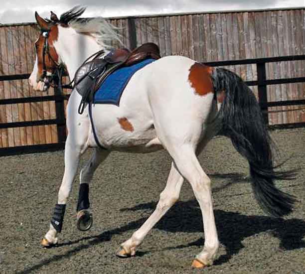
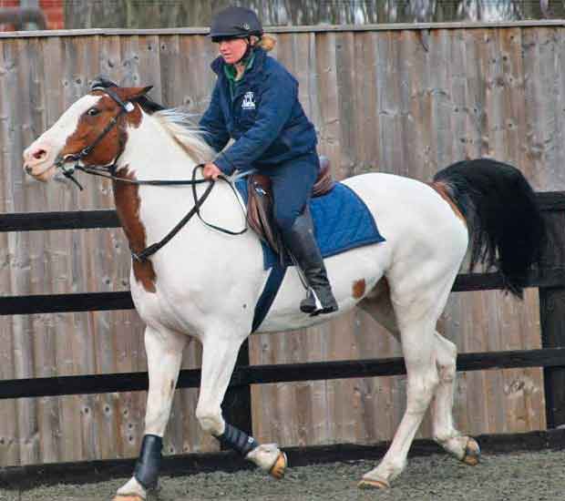
If the rider perceives a pain-related issue is affecting the horse’s ridden performance, it is hugely important the horse is evaluated ridden (Figures 1 and 2), because some gait abnormalities are only apparent when the horse is ridden (for example, those related to sacroiliac joint region pain; Barstow and Dyson, 2015). Equally, lameness seen in hand or on the lunge may not be the only, or even the main, contributor to musculoskeletal pain experienced by the horse when it is ridden.
Poor agreement exists among veterinary surgeons for lameness identification, particularly for hindlimb lameness, and low-grade lameness can be especially difficult to identify (Keegan et al, 2010). Lack of visual movement asymmetry (“head nod” for forelimb lameness and “hip hike” for hindlimb lameness) does not preclude the presence of pain.
Logically, bilaterally symmetrical lameness will not result in movement asymmetry, and may be manifest as shortening of the step length or a lack of impulsion. In addition to movement asymmetry, other indicators of musculoskeletal pain include the presence of saddle slip (Greve and Dyson, 2013), bucking and gait abnormalities in canter (Barstow and Dyson, 2015) and asymmetrical body lean angles between the left and right reins (Greve et al, 2018).
A body of research has shown certain facial expressions and behavioural signs may reflect musculoskeletal pain (Dyson et al, 2017; Mullard et al, 2017), and a ridden horse ethogram has been developed for the detection of musculoskeletal pain (Dyson et al, 2018). Potentially, this is a very useful tool for identification of pain-related problems in ridden horses.
If behavioural indicators of pain are identified in the absence of overt lameness, referral may be the best option, because the horse may require evaluation across several days. Another method of determining whether the problems perceived by the rider are pain related is to assess the response to analgesic drugs such as phenylbutazone.
Improvement of clinical signs during the trial period, followed by deterioration after analgesia is withdrawn, indicates a probable pain-related problem. False-negative results are possible if an inadequate dose or length of administration is used; phenylbutazone, 4.4mg/kg per os bid, administered over a minimum of seven days, is recommended.
In addition, certain conditions, such as sacroiliac joint region pain, are known to often be non-responsive to oral analgesia; therefore, a negative response to an analgesic trial does not preclude the presence of a pain-related problem. Although interpretation relies on the rider, who may be prone to the placebo effect, a positive response is generally a reliable indicator of a pain-related problem and prompts further investigation.
A thorough and systematic approach to the physical examination of the whole horse is essential in all cases to ensure important information is not missed. Owners may voice their opinions as to where they believe problems are. However, it is important not to be swayed by these. They may be incorrect, and clinical signs are commonly present in areas distant to the underlying source of pain, such as thoracolumbar stiffness (decreased range of movement) secondary to hindlimb lameness (Greve et al, 2017), or epaxial muscle tension or pain secondary to sacroiliac joint region pain (Barstow and Dyson, 2015).
The physical examination would ideally be performed in the stable so the horse is relaxed. Veterinary surgeons should develop, and strictly follow, a methodical approach to visual appraisal and the palpation of each structure. This should include palpation of the limbs in both weight-bearing and non-weight-bearing positions, manipulation of the joints, and palpation of the major muscle groups for tension or pain.
The flexibility and stability of the cervical and thoracolumbosacral spine should be assessed. Attention should be paid to the horse’s reactions to palpation and manipulation. In-depth knowledge of musculoskeletal anatomy is essential, as is the knowledge of what is normal versus abnormal, and whether abnormalities are likely to be of clinical significance.
Cobs with hairy legs and/or thick, folded skin pose a particular challenge. Removal (clipping) of long hair may be the only solution to allow physical examination of the lower limbs, and if diagnostic analgesia is to be performed, it is essential.
Swellings or distension of a joint or thecal capsule accompanied by heat and/or a painful response to palpation or manipulation are likely to be clinically significant. However, it is important to complete the full physical and gait examinations and to confirm the location of all sources of pain contributing to lameness (unless the history and severity of lameness is indicative of a possible fracture), because additional sources of pain may exist.
Thoracolumbar stiffness and epaxial muscle tension and pain are commonly secondary to distant sources of pain, rather than indicative primary thoracolumbar pathology (Zimmerman et al, 2011). Impinging spinous processes (so-called “kissing spines”) do not necessarily always cause pain. The presence of impinging spinous processes in a horse with clinical signs of pain relating to the back can be a red herring in our experience, and diagnostic analgesia is always required to confirm or rule out impinging spinous processes as a source of pain.
Visual appraisal should be performed from all sides of the horse, with it standing squarely and weight bearing evenly on all limbs on a flat surface, preferably outside. The hindquarters should be assessed for symmetrical or asymmetrical development of the gluteal or quadriceps muscle groups, and for relative heights of the tubera sacrale and tubera coxae.
Chronic hindlimb lameness can lead to disuse atrophy of the hindquarter muscles. Left-right asymmetry of the tubera sacrale or coxae or gluteal musculature is important to identify prior to gait evaluation, to avoid misinterpretation of physical hindquarter asymmetry as movement asymmetry.
Assessment of conformation is still largely based on empirical knowledge. However, several conformational traits may be associated with certain injuries: straight hindlimbs (large hock angles) have been associated with proximal suspensory desmopathy (Dyson and Murray, 2011; Routh et al, 2017); tubera sacrale higher than the withers may be associated with sacroiliac joint region pain (Dyson and Murray, 2003); and horses with poor foot conformation are at risk of foot-related problems (Holroyd et al, 2013).
Gait evaluation is essential for the identification of lameness and poor performance, and localisation of the source of pain, through assessment of the response to diagnostic analgesia. Adequate facilities and a competent handler are essential prerequisites.
Horses should first be seen moving in hand on a hard surface. It is important the surface is even, and long enough to allow the horse to trot at a steady speed before having to stop. Not all yards have ideal facilities to allow this, and performing the trot-up on roads can be potentially hazardous.
Unless lameness is marked and further evaluation is unwarranted, evaluation under additional circumstances, including lungeing on soft and firm surfaces and ridden exercise, should be performed. As a result of sometimes non-ideal facilities at a client’s premises, this is not always possible in first-opinion practice.
Several objective gait analysis systems are readily available to veterinary surgeons. Growing evidence suggests inertial measurement units (IMUs) can be used to accurately quantify movement of the head and tubera coxae and/or tubera sacrale, and aid in the detection of mild lameness (Pfau et al, 2014, Rhodin et al, 2018). They can also be used to provide some unbiased objective data for the assessment of response to diagnostic analgesia (Pfau et al, 2014).
While these systems are useful tools for the identification of unilateral lameness and in the assessment of gait alteration after diagnostic analgesia, users must be aware of several shortfalls of these systems. Firstly, these systems identify asymmetry of movement, and so bilaterally symmetrical forelimb or hindlimb lameness without movement asymmetry may not be identified. Another major limitation is not enough is known about the movement patterns of non-lame and lame horses on the lunge and when ridden, so these systems cannot currently be reliably used under these circumstances.
Movement asymmetry is only one gait adaptation employed by horses to avoid pain. Many other gait adaptations indicative of lameness can be evaluated, including reduced step length, reduced height of arc of limb flight ± toe drag, reduced or increased step frequency and altered limb placement (moving crookedly). Objective gait analysis may aid practitioners in the detection of subtle unilateral lameness; however the limitations of these systems are significant and they should always be used judiciously and in conjunction with subjective evaluation.
Diagnostic analgesia (nerve blocking) is crucial for determining the source of pain causing the lameness. In the absence of localising clinical signs it is the gold standard procedure for the localisation of pain. The majority of diagnostic analgesic techniques can be performed relatively easily; however, this is a potentially hazardous procedure and a competent handler is essential.
Horses that are potentially dangerous when performing nerve blocks are usually good candidates for referral. Experienced staff, and facilities such as wooden stocks for hindlimb blocking, are invaluable for performing nerve blocks in fractious horses.
A minority of horses may require heavy sedation, which prolongs the investigation and is, therefore, unfeasible for first opinion practitioners. Nerve blocking horses with multi-limb lameness can also be a lengthy procedure.
As well as being potentially problematic for the typically busy first opinion practitioner, this can be costly for the owner and referral may be more economical because several investigations can be performed alongside each other at a clinic facility, leading to lower costs.
A growing body of evidence suggests perineural analgesia is not as specific as once thought, and results can sometimes be misleading. Diffusion or incorrect deposition of local anaesthetic solution can desensitise regions proximal to the site of injection (Nagy et al, 2009; Nagy and Malton, 2015) or adjacent structures (Dyson et al, 2004; Nagy et al, 2012; Contino et al, 2015). Accuracy of needle placement and use of small volumes of local anaesthetic solution may improve specificity compared with the use of larger volumes, which may diffuse further.
Intra-articular blocks are more specific, although they may still desensitise adjacent nerves. For example, intra-articular analgesia of the tarsometatarsal joint may anaesthetise the deep branch of the lateral plantar nerve (Contino et al, 2015). Therefore, cross-blocking on a separate occasion is often required to try to definitively ascertain the location of pain. Nonetheless, this potential lack of specificity must be borne in mind when performing radiography and ultrasonography.
With intra-articular injection (and perineural injections performed in close proximity to joint capsules) comes the low risk of joint sepsis (Bohlin, 2014). In first opinion practice this risk may be higher because of the increased potential for contamination in an uncontrolled environment. With infiltration of local anaesthetic solution around the sacroiliac joint regions a low risk of transient femoral nerve paralysis is possible and, rarely, temporary recumbency.
Diffusion is probably influenced by injection technique, volume used, and individual anatomic variation. Adequate management of an ataxic or recumbent horse over several hours is often not possible in first opinion practice, because of a lack of facilities at yards, lack of skilled handlers, and lack of time available for constant monitoring by the veterinary surgeon. Horses displaying the typical clinical signs of sacroiliac joint region pain (Barstow and Dyson, 2015) are, therefore, good candidates for referral.
Screening evaluations using skeletal scintigraphy (bone scan) were historically used for sports and leisure horses with complex or multi-limb lameness, poor performance, lameness judged to be too mild to reliably interpret the results of nerve blocks and horses in which performing nerve blocks is judged to be hazardous. However, a recent large-scale study of 480 sports and leisure horses with lameness or poor performance reported that when skeletal scintigraphy was used as an indiscriminate screening evaluation, the results were unlikely to lead to a full and correct diagnosis of the underlying cause(s) of musculoskeletal pain (Quiney et al, 2018).
The overall specificity was high (94%), but the sensitivity was low (43.8%), meaning a high probability of false-negative results that could, in turn, lead to missed diagnoses. The greatest proportion of false-positive results occurred at the distal aspect of the hock (27.2%) and the sacroiliac joint region (12.5%).
Increased radiopharmaceutical uptake in these regions should, therefore, be interpreted with caution, to avoid misdiagnosis. Little value is seen in radiographic investigation or presumptive treatment based on the presence of increased radiopharmaceutical uptake, unless pain has been localised to the region.
![Figure 3. [A] Dorsoplantar radiograph of a hock acquired with a horizontal beam. The centrodistal and tarsometatarsal joint spaces are poorly defined (arrows). Artefactual narrowing of the distal tarsal joints is common in dorsoplantar images acquired with a horizontal beam because the distal tarsal joints slope from proximal, dorsally to distal, plantarly. [B] Dorsoplantar radiograph of the same hock as in A, acquired with a beam angled 5° distally (dorsal 5° proximal-plantarodistal oblique). The centrodistal and tarsometatarsal joint spaces are clearly visible and no narrowing exists. [C] Lateromedial radiograph of a foot of non-diagnostic quality. This is not a true lateromedial because the distal condyles of the middle phalanx and the palmar processes of the distal phalanx are not superimposed. The demarcation between the palmar compact bone of the navicular bone and the spongiosa, and the integrity of the palmar compact bone, cannot be assessed, because the beam is not perpendicular to the long axis of the navicular bone. The navicular bone cannot be evaluated using this image. [D] Lateromedial radiograph of the same foot as in C, acquired with better positioning of the foot. This image is of good diagnostic quality. The palmar compact bone of the navicular bone is well demarcated because the beam is perpendicular to the long axis of the navicular bone. There is mild proximal and distal extension of the palmar compact bone of the navicular bone.](https://www.vettimes.co.uk/app/uploads/2019/03/VTE5.1_Quiney_Dyson_Figure-3_feat.jpg)
[B] Dorsoplantar radiograph of the same hock as in A, acquired with a beam angled 5° distally (dorsal 5° proximal-plantarodistal oblique). The centrodistal and tarsometatarsal joint spaces are clearly visible and no narrowing exists.
[C] Lateromedial radiograph of a foot of non-diagnostic quality. This is not a true lateromedial because the distal condyles of the middle phalanx and the palmar processes of the distal phalanx are not superimposed. The demarcation between the palmar compact bone of the navicular bone and the spongiosa, and the integrity of the palmar compact bone, cannot be assessed, because the beam is not perpendicular to the long axis of the navicular bone. The navicular bone cannot be evaluated using this image.
[D] Lateromedial radiograph of the same foot as in C, acquired with better positioning of the foot. This image is of good diagnostic quality. The palmar compact bone of the navicular bone is well demarcated because the beam is perpendicular to the long axis of the navicular bone. There is mild proximal and distal extension of the palmar compact bone of the navicular bone.
These systems can produce images of excellent quality, quickly, at the horse-side. Digital storage systems (picture archiving and communication system) are essential for archiving images, and make the communication of images for second, or expert, opinion possible. However, the quality of interpretation cannot exceed the quality of image acquired. Image quality largely depends on user knowledge and experience, and requires precision.
In addition to the selection of appropriate exposure factors, appropriate beam centring and collimation, and patient preparation and positioning can vastly alter image quality (Figure 3). Each image should be critically appraised for diagnostic quality, and repeated if necessary.
If the cause of pain cannot be identified from conventional imaging, or if the abnormalities identified do not correlate with the clinical picture, referral for advanced imaging, such as MRI or CT, can provide additional information.
Lameness investigation is both a science and an art, and with a logical, thorough and judicious approach, high-quality lameness evaluations can be performed in first opinion practice – facilities and competent handler availability permitting. However, the investigation of lameness without obvious localising clinical signs or non-specific poor performance can be very time-consuming and is often not best suited to first opinion practice.
The interpretation of the response to diagnostic analgesia is not always straightforward and must be done in light of the clinical picture and history – this requires experience that is not acquired through university education. Competence will come with experience, and referral of cases for lameness investigation can be of huge learning benefit for inexperienced and experienced referring veterinary surgeons.
Prescribing or administering any treatments without a definitive diagnosis or a clinical indication is ill-advised, because this is often a waste of client money, may be of no benefit to the horse or even contribute to lesion deterioration, increases time out of work, leads to a poorer outcome, and negatively impacts on client trust.
Equally, performing diagnostic imaging without localising the source of pain is inappropriate and may lead to misdiagnosis. In the absence of obvious localising clinical signs, what can be reasonably done in first opinion practice is relatively limited. The importance of early recognition of pain-related problems in the ridden horse cannot be stressed enough.
With the poor abilities of many riders, owners and trainers to identify the behavioural signs of pain that compromises the welfare of a disturbingly large proportion of horses in ridden work, arguably, one of the biggest impacts the first opinion practitioner can make on equine welfare is to identify these horses and help educate owners in the recognition of pain.
Our ability to diagnose musculoskeletal pathology currently exceeds our ability to satisfactorily treat it. The ability to recognise and manage lameness earlier than we have done historically may be our best armament against the most common condition affecting horses.