2 May 2016
Jonathan Anderson discusses the range of tendon injuries that can lead to a loss of potential in performance horses, as well as various therapies that help reduce recovery time.
Jonathan Anderson
Job Title
Figure 4. The use of underwater treadmill therapy offers the advantages of cold therapy in a controlled exercising regime tailored according to the injury and stage of rehabilitation.
Tendon injuries in performance horses range from career-ending breakdown injuries in Thoroughbred racehorses to low grade, but persistent, injuries of the deep digital flexor tendon (DDFT) in the foot of dressage horses.
Both are career-limiting, have expensive and long-term consequences for the athletic future of the horses, and can result in disappointment and heartache for the owner and his or her connections. Tendon injuries are a serious cause of loss of athletic potential, result in considerable time and economic cost, and require careful diagnosis with regular veterinary input to assess their healing and appropriate management.

Tendons are divided into flexor tendons and extensor tendons by virtue of their function on the limb.
Tendons are composed of collagen fibres grouped in increasingly larger subunits divided by looser connective tissue (endotenon; Figure 1). These fascicles slide over each other by virtue of the endotenon and have a waveform known as crimp.
The endotenon is continuous with the epitenon – the layer of connective tissue surrounding the outside of the tendon. In tendons not contained in a sheath, the thicker paratenon circumferentially wraps around the entire unit of tendon fibres. The paratenon stretches considerably, so is usually not ruptured when a tendon does. It provides vascularity and cellular elements and, therefore, is important for repair. The lack of it in tendons in sheaths is one reason for poor healing.
Tendons are 70% water and 30% type one collagen. The structural and material properties of a tendon depend on the spatial organisation of the collagen fibrils and their cross linking.
Cartilage oligomatrix protein (COMP) is a non-collagenous protein that organises the fibril framework during formation and growth, and increases dramatically, peaking at two years. When tendons pass over joints, they are usually contained within a sheath, providing a synovial environment for smooth gliding.
Tendons are highly elastic and confer a tremendous amount of support for the appendicular skeleton. They are highly exposed and susceptible to injury, either from excessive strain or external trauma due to their proximity to the surface of skin. Except for wire injuries, lacerations to the tendons usually occur when they are under the greatest weight bearing load and up to 50% of the tendon can be lacerated without compromising the horse’s ability to ambulate.
Tendon injuries can occur in two broad ways.

Overstrain occurs either from a sudden overloading of the structure – typical for DDFT injuries – or strain-induced as a result of a preceding phase of molecular degeneration (inflammation), which progressively weakens the tendon. This is evidenced by lesions in “normal” tendons, the increase in blood flow in some tendons, and the relationship between injury and increasing age and intensity of exercise.
The “core lesion” in the horse is likely a result of decreased collagen crimping in the centre of the tendon compared to the surrounding fibres, resulting in the central fibres being straightened maximally first.
The superficial digital flexor tendon (SDFT) can adapt to exercise during skeletal development, but after skeletal maturity the synergistic effect of ageing and exercise causes degeneration and cumulative microdamage (Smith et al, 2002). As the peak load exceeds the tendon’s structural strength, physical disruption from fibrillar slippage, breakage of cross-linking elements to fibrillar rupture and tendon disruption occur (Avella et al, 2009).
Tendons are viscoelastic and can stretch 10% to 12% of their length before they rupture in vivo. In vitro normal strains in digital flexor tendons in vivo are 3% to 8% at walk, 7% to 10% at trot and 12% to 16% at gallop (Stephens et al, 1989).
Tendons transmit a great deal of energy between loading and unloading in the form of heat, which is responsible for rises in temperature as high as 44% in the centre, leading to the hypothesis damage to the tendon matrix is related to core temperature increase (Avella et al, 2009).
The discipline of the horse often dictates the type of injury. SDFT injuries are seen commonly in disciplines involving fast work, such as event horses, and Thoroughbred and standardbred racehorses. DDFT injuries are commonly seen in other sport disciplines, such as showjumping, endurance and dressage.
Risk factors increasing the load on SDFT include increasing speed of the horse, age, a firmer track surface, weight being carried and hoof conformation. For the latter, the classic long toe, low heel front foot conformation favoured by many Thoroughbred trainers has the effect of increasing the angulation of the metacarpophalangeal (MCP) joint and places greater strain on the SDFT, increasing the damage risk.
There is a protective effect of an increasing heel angle for DDFT; however, caution needs to be used as it results in extension of the fetlock that can place greater strain on the suspensory ligament and SDFT.
The SDFT extends from the palmar aspect of the carpus, around medial to the accessory carpal bone and extends distally on the midline down the palmar aspect of the third metacarpal bone (MC3), over the MCP joint and into the pastern region. Starting off as an oval structure, it becomes more banana-shaped as it extends distally in the metacarpal region and becomes a flattened banana-shaped structure that circumferentially wraps around the DDFT in the digital tendon sheath (DTS)by virtue of the thin fin-like extension of the manic flexora. The SDFT is enclosed in the DTS from the mid to distal third of the MC3 to the distal pastern region. At the second phalanx (P2) level it branches into lateral and medial branches to wrap around the P2 to insert on to the dorsal aspect.
In the metacarpal region, as the most superficial structure lying directly beneath the skin and surrounded by paratenon that provides its blood supply and nutritional support, it is susceptible to traumatic injury and lacerations in the palmar metacarpal region will often involve variable degrees of SDFT damage.
The SDFT proximal to the mid-MC3 region is the most common site of injury. As the heel strikes the ground the loads rise most quickly in the soft tissue structures supporting the MCP joint – namely the SDFT and the suspensory ligament. The load in the DDFT rises later, so is less frequently injured.
In the tendon sheath, the DDFT is more commonly injured in the forelimb, the foot and the pastern region, although branches of the SDFT can sustain injury, resulting in lameness.
The manica flexoria is a common site of injury in the hindlimb of sports horses, with fibrillation and tears resulting in tendon sheath effusion and mild to moderate lameness. Lesions of the manica flexoria are more common in hindlimbs than forelimbs due to getting caught as it passes through the fetlock canal.
Tears result in tenosynovitis due to the inflammatory response created by the torn tendon. Ultrasonographically, non-specific findings, such as thickening of the mesotenon of the DDFT, tenosynovitis and blunting of the SDFT, can accompany tears of the manica flexoria.

Visualisation of the manica flexoria specifically is difficult given its close proximity to the DDFT. Contrast radiography, MRI and CT of the DTS can help in diagnosis of manica flexoria tears, but tenoscopic evaluation is the most accurate in allowing direct visualisation, as well as surgical debridement. This is necessary to allow debridement of tears and fibrillation and, in most cases, resection of the manica flexoria.
Core lesions of the SDFT in horses occur in all disciplines, but are most commonly seen in Thoroughbreds. Such lesions reflect a combination of strain and stretch, and thermal insult, resulting in fibre degeneration and creation of a haemorrhagic lesion.
Core lesions result in marked lameness that can quickly resolve over hours. As with any soft tissue trauma, tendinous injury results in initial intratendinous haemorrhage, followed by a pronounced inflammatory reaction, oedema and swelling of the limb. These have a detrimental effect on the tendon, leading to greater damage.
The inflammatory phase rapidly overlaps in the reparative phase of healing, where there is a pronounced angiogenic and fibroblastic response, resulting in the formation of collagen type III, typical of scar tissue. This is inferior to the type I collagen of normal tendon fibres.
The reparative phase overlaps in the remodelling phase of healing, during which there is a gradual, but incomplete, transformation to type I collagen as scar tissue matures. The scar tissue is stiffer and the repaired tendon is strong, but is functionally inferior to normal tendon, so is predisposed to re-injury at sites adjacent to the original injury. The absence of a paratenon in tendons intrathecally results in an efficient repair.
Clinically, tendon lesions usually result in palpable heat, pain and swelling at the site of injury, although, in many cases, this is not evident in the initial stages and are often present in contralateral limbs.
Observable lameness is often marked initially, but may resolve quickly and is related to the degree of inflammation more than the degree of damage. Once the inflammatory phase passes, lameness typically resolves. Careful palpation of each tendinous structure is essential with the limb in weight bearing and non-weight bearing positions.
Pain response differing on each side and careful visual assessment can also help identify the region of injury. Typical “bowing” of the limb develops during 7 days to 14 days and is a very identifiable indicator of an insult to the underlying tendon (Figure 2).

Lesions in the SDFT can extend the entirety of its length; therefore, it is important to assess the SDFT in the palmar aspect and proximal to the carpus, as well as distal to the carpus. Lesions extending proximal to the carpus carry a guarded prognosis traditionally although – with an increased use of water-based rehabilitation therapies and biologic preparations – better results have been achieved, in the author’s experience.
Ultrasound is the most useful tool to evaluate the tendon, and Doppler machines help determine the vascularity and chronicity of the lesion, as well as differentiating inflammatory granulation tissue, as apposed to scar tissue in chronic lesions (Kristofersson et al, 2005).
Both longitudinal and transverse views of the tendon should be obtained and the limb should be clipped. For Doppler use, the tendons are scanned in the relaxed position without a standoff for 30 seconds. A standoff is essential to detecting irregularity of the palmar surface of the tendon, as well as superficial lesions of the SDFT. It is also advisable to ultrasound the contralateral limb, as approximately a third of strain-induced tendon injuries will have bilateral components (Avella et al, 2009).
Ultrasound can be performed any time post-injury, but a more accurate assessment of lesions is best seen after the inflammatory phase of injury has passed – 7 days to 10 days post-injury. Changes in shape, size, position and echogenicity are assessed along with the irregular striated fibre pattern typical of fibrosis in chronic tendinopathies.
Diagnosis of tendon pathology in the pastern and foot has been enhanced by the advent of contrast enhanced CT and MRI, both providing multiplanar slices of detailed anatomical information in three different planes (Figure 3). This has allowed characterisation of specific types of lesions affecting the insertion and portion of the DDFT adjacent to and overlying the navicular bone.
The DDFT is most frequently injured in the DFTS or adjacent to, or at the level of, the distal sesamoidean bursa (Blunden et al, 2009). Intrasubstance tears that may have burst out the lateral and medial borders of the tendon are most commonly seen at the level of the collateral sesamoidean ligament and the distal sesamoidean bone.
CT with intra-arterial contrast has also been used to successfully document tendinopathies in the hoof and tendon sheath. Often, these are not ultrasonographically visible. The use of contrast enhanced tenographic CT has been documented as an aid in diagnosis of recognition of manica flexoria tears in the hindlimbs of horses (Lacitignola et al, 2015). Studies are ongoing to determine the accuracy of the technique in the forelimbs.
Thermographic imaging of the distal limb is fraught with interpretation difficulties relating to fluctuations in ambient and environmental temperatures. However, correcting for these differences, serial measurements of the same horse may help to detect the effect of early injury to the limb before any swelling or observable lameness is recognised.
Although the use of biological markers has not led to the production of a clinically accessible test to detect tendon damage, several proteins have shown promise. Significant increases in COMP in the DTS synovial fluid of disrupted tendons made it a potential biomarker to predict early tendon damage (Smith et al, 2011).
In older horses, increased COMP levels with DTS synovial fluid was strongly suggestive of a tear, despite lack of ultrasonographic evidence of a lesion. Synovial fluid analysis may provide the future for detecting early tendon pathology; however, it is not available as a routine test.
Treatment of acute tendon lesions requires the use of cold therapy, rest, anti-inflammatory and analgesic support, as well as compressive and supportive bandaging techniques to reduce the formation of inflammatory mediators, decrease enzymatic activity, slow down nerve conduction and reduce oedema formation.
Cold hosing of 20 minutes several times daily, the use of ice and vibration devices, and underwater spa therapy have all been used successfully in acute phase management. Cold hydrotherapy is superior to ice packs because of increased contact and it does not cause freeze burns (Petrov et al, 2003).
Cold therapy should not be applied for longer than 30 minutes and temperatures below 0°C are not advisable. Cold water spa therapy provides cold and compression with hypertonic saline at 5°C to 9°C and is increasingly used in rehabilitation programmes in which therapy can be tailored for the horse and injury (Hunt, 2001; Figure 4).
Compression reduces inflammation and oedema by increasing interstitial hydrostatic pressure. A modified Robert Jones bandage is suitable in most cases, with a palmar/plantar splint or cast applied where hyperextension of the fetlock is evident. The contralateral limb will provide a good template for shaping a splint.
NSAID and corticosteroid administration in the first 24 hours to 48 hours following injury are beneficial. Both provide analgesic relief in the initial stages following injury and corticosteroids are a potent anti-inflammatory. After 48 hours, the use of corticosteroids is not recommended due to the inhibitory effects on fibroplasia. Dexamethasone (0.1mg/kg) as a single dose is recommended as it has little risk of laminitis.

Confinement and cessation of exercise, followed by a controlled exercise regime, form the most important part of a tendon’s ability to heal. It helps to resolve inflammation and promote optimal collagen remodelling. Injuries of the SDFT require at least 9 months to 12 months of rehabilitation before the resumption of full athletic function.
Rehabilitation longer than 18 months is unnecessary as healing appears complete by this stage (Dyson, 2004). Rehabilitation programmes are designed around the ultrasonographic and physical characteristics of the lesion and it is important to document the progress of healing ultrasonographically and tailor the rehabilitation programme accordingly.
Measurement of the cross-sectional area allows assessment of size and increases of 10% between examinations suggests a degree of re-injury requiring a change in the implemented exercise regime. Ultrasound appearance will be unlikely to alter significantly before eight weeks once the inflammatory phase of healing has been completed, and changes in intensity of exercise should be monitored closely for how the tendon is adapting to strain.
Uncontrolled turn out exercise is detrimental to the horse’s return to athletic function, with one study showing a controlled exercise regime resulting in 71% of horses returning to athletic function versus 25% in an uncontrolled programme (Gillis, 1997).
Therapeutic ultrasound, laser and magnetic devices are controversial in their beneficial effects on tendinopathic healing. Ultrasound likely works in the conversion of sound to thermal energy that has been shown to increase vascularisation and fibroblastic proliferation compared with controls (Morcos and Aswad, 1978). Low-level lasers stimulate metabolism and enhance fibroblastic proliferation, but no data demonstrates the effect on tendon healing. Magnetic therapy has not been documented as beneficial for tendon healing in any clinical trial, despite its widespread use by owners.
Counter-irritation continues to be used in some areas to increase the formation of fibrous scar tissue around, and in regions of, tendon damage.

Topical iodine-based and mercury-based compounds, intralesional hyalase and thermal cauterisation using heated bars or pins applied on the skin over which the tendon is damaged or into the lesion, have been used in an attempt to increase the fibrous scar tissue reaction in and around the tendon.
This does not result in a stronger tendon, with beneficial effects likely attributable to the enforced rest period following the procedure. Therefore, its use is controversial.
A range of medial and surgical treatment modalities has been described for superficial flexor tendon lesions. Most involve the use of rest and remedial farrier work, in conjunction with some specific intralesional therapy.
Intralesional therapeutics are injected under ultrasound guidance in the standing, sedated horse, with the limb blocked locally or regionally and following strict aseptic techniques (Figure 5). A minimum period of three days post-injury is required, so as not to increase haemorrhage intralesionally. The volume used should be based on the size of the lesion with “over filling” of lesions resulting in more damage.
Platelet-rich plasma (PRP; Figure 6) and bone marrow derived from mesenchymal stem cells (MSCs) are most popularly used; however, polysulphated glycosaminoglycans, hyaluronic acid (HA) and non-depot formulations of corticosteroids have shown variable degrees of efficacy for tendon healing in reducing inflammation (Fowland et al, 1992; Redding et al, 1992).
HA administered intrathecally reduces adhesion formation in the DTS following tendinopathy and associated tenosynovitis. The carrier in which methylprednisolone acetate is formulated results in dystrophic mineralisation and tissue necrosis when injected via intratendinous and peritendinous means, and should not be used.
PRP is a rich source of growth factors that stimulate cell proliferation and matrix synthesis (Bosch et al, 2010).
Although long-term clinical data is lacking, it is widely used for both chronic and acute tendon lesions and shows promising efficacy in comparison to other similar treatment formulations.
Bone marrow and fat-derived MSCs induce a matrix more closely resembling normal tendon than the fibrous scar tissue formed by natural repair, and are used safely and extensively to treat lesions in both tendons and ligaments of equines.
Pluripotent cells have the potential to differentiate into tenocytes, thereby providing a source of true tissue regeneration that results in a superior tendon construct than scar tissue fibrosis.
Following treatment, reinjury rate in national hunt racehorses was 26%, with more than three years of follow-up, which was significantly better than for horses treated and analysed conventionally (Godwin et al, 2012).
The combination of PRP and MSCs has shown to result in decreased inflammation and greater organisation than saline-treated controls in a controlled trial (Carvalho et al, 2013). The use of MSCs provides the greatest promise of enhancing repair; however, the signals that determine direction of differentiation from pluripotent stem cell to tenocyte remain little understood.

Basic surgical intervention includes decompression of a lesion using needles or a number 11 scalpel blade (tendon splitting) with or without intralesional therapy. First described in the 1940s, it is used to decompress the acute anechoic core lesion, facilitates vascular in growth and may reduce the lengthening of the lesion (Henninger et al, 1992). Bandaging and resting for two weeks is required prior to a controlled exercise regime.
Desmotomy of the superior check ligament (accessory ligament of the SDFT) is designed to take the strain off the SDFT by producing a functionally longer musculotendinous unit; however, in equine cadaver models, the procedure increased strain on the SDFT due to extension of the MCP joint.
In vitro, other biomechanical alterations are likely at play – resulting in the success of the procedure reported in racehorses (Gibson et al, 1997). The procedure can be performed via tenoscopy and is reserved for horses with repeat SDFT lesions or extensive lesions that extend proximal to the carpus.
Intrathecal tears of the DDFT – in the navicular bursa and DTS – and the manica flexoria component of the SDFT are best-managed via tenoscopy by debridement or, in the case of the manica flexoria, by removal. Tears of these structures commonly result in effusion of the sheath, which often fails to resolve following debridement (Figure 7). Dorsal tears and fibrillation of the DDFT in the bursal region are identified by MRI and can be debrided via bursoscopy.
Lacerations of specific tendons result in characteristic clinical presentations and SDFT lacerations result in hyperextended (dropped) MCP joint when the limb is loaded.
If both SDFT and DDFT are lacerated, the toe and hyperextended fetlock joint will raise from the ground. Extensor tendon lacerations, most commonly in the mid-metacarpal/metatarsal region, result in occasional stumbling on to the dorsal aspect of the fetlock joint and an inability to protract the limb completely. They rarely result in long-term gait abnormalities or lameness.
As with any laceration, the proximity to synovial structures is essential as the secondary septic synovial process is likely to be more detrimental to the horse than the lacerated tendon.
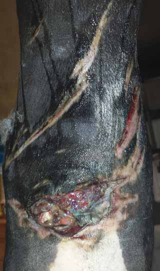
Frequently, extensor tendon sheath lacerations can go undetected until there is considerable swelling of the dorsal aspect of the limb, at which time, treatment becomes orientated around the management of a chronic sepsis than an injury to the tendon itself. Ultrasound is essential to determine the synovial and tendinous structures involved (Figure 8).
As acute transection of the flexor tendons is most commonly associated with open skin lacerations, it requires emergency surgery to debride and, in some cases, suture the tendon ends. Most commonly, the ability to suture the ends of the tendon is made difficult due to the recoil of the ends proximal and distal to the wound, as well as the inevitable contamination that accompanies the open wound. Following debridement, autologous grafts can be used to bridge the gap and provide a scaffold for new tendon fibres to grow across.
These injuries require supportive bandaging and casting with a heel elevation incorporated in the cast to prevent the fetlock from dropping and allow as close an apposition as possible of the tendon ends. Lacerations involving 50% or less of the tendon require debridement only.
Casting is required for a minimum of six weeks to eight weeks, by which point the breaking strength of the tendon is the horse’s bodyweight. Stall rest of four months is required before exercise is resumed and a year is needed before return to athletic function.
It is important on cast removal the heel elevation is removed, as this results in extension of the fetlock and leads to excessive strain on the SDFT. Less commonly, swelling on the lateral aspect of the carpus is associated with carpal sheath tenosynovitis – most commonly associated with DDFT injury. Arthroscopic debridement and controlled exercise regimes are necessary for healing, although swelling of the sheath often persists.
In the hindlimb, the SDFT flattens out over the plantar aspect of the tuber calcanei and stabilised by tight medial and lateral retinacular bands that attach to the medial and lateral aspects of the tuber calcis. At this point, the tendon is composed of fibrocartilage and tendinous fibres.
Direct trauma or excessive strain during partial flexion can cause, in decreasing frequency, lateral or medial luxation, or splitting of the SDFT, with two parts sitting either side of the tuber calcis. It results in acute swelling and usually luxates on and off the tube calcis, depending on the weight-bearing stance of the horse, and a marked gait abnormality with variable degrees of distress.
Ultrasound can help in determining the position of the luxation when the swelling makes it difficult to visualise and palpate. Partial or intermittent luxations can heal with stall rest, with or without a bandage or full limb cast, but rest for four months to six months is necessary.
Surgical repair is considered mandatory for a return to athletic function, although the conversion of partial luxation to complete luxation by transection of the apposing retinaculum can also return horses to athletic capabilities. Surgical repair involves meshes or screws to fix the torn retinaculum to tuber calcis and requires full limb casting for recovery and a four month to six month period of stall confinement to achieve adequate fibrosis. Medial dislocations have a better prognosis than lateral displacement; however, conservative and surgical treatments carry a guarded prognosis for athletic return.
Spontaneous rupture of the SDFT without the presence of a wound is recognised in older horses. These are managed with minimal supportive bandaging – often healing after four months to six months of confinement and six months of controlled exercise.
On the dorsal aspect of the forelimb the common extensor tendon extends from its muscular counterpart proximal to the carpus, extending on the dorsal aspect of MC3 to insert into the mid to distal P2. The tendon is enclosed in a tendon sheath over the dorsal carpus and is a relatively frequent site of injury due to its position directly beneath the skin.
Typically, a puncture or laceration will result in damage to the tendon sheath and tendon, requiring surgical management in the form of tenoscopic-assisted lavage of the sheath under general anaesthesia.
Rupture of the common digital extensor (CDE) tendon is an occasional result of laceration on the dorsal aspect of the MC3 and leads to initial marked lameness that can quickly progress to relatively minor lameness. The horse learns to adapt to the inability to extend the toe by flicking the foot forward prior to placement on the ground surface. Such lacerations are rarely a cause of long-term concern.
The extensor carpi radialis extends lateral to the CDE on the dorsal aspect of MC3 and has a sheath as it extends over the dorsal aspect of the carpus. Injury to this tendon, usually after penetration of the sheath, requires lavage and debridement under tenoscopic visualisation.
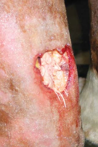
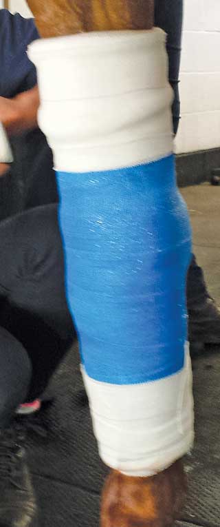
Extensor tendon lacerations heal very well without tenorrhaphy and often require wound (Figure 9) and sheath management, and six weeks of stall rest for healing. Casting of the limb may help in cases where there is extensive soft tissue contusion (Figure 10). In chronic cases of CDE-septic tenosynovitis non-responsive to treatment, removal of the sheath and CDE can result in a successful return to exercise (Booth et al, 2004).
The prognosis for tendon injuries varies depending on its location, the involvement of synovial structures, the presence of wounds and the level of expected athleticism, and is unknown until treatment is underway.
Published studies can aid in prognostication: