28 Nov 2016
Patrick Pollock discusses a development in equine veterinary medicine that has led to a fundamental change in understanding the airway – both in terms of health and disease.
Patrick Pollock
Job Title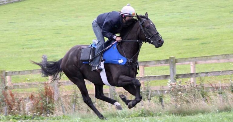
Figure 1. A horse undergoing overground endoscopy with its usual rider on its home gallop.
The most frequently reported consequences of upper respiratory tract obstruction in equine athletes are poor performance and abnormal respiratory noise (Morris and Seeherman, 1991; Martin et al, 2000). However, diagnosing the cause of such non-specific clinical signs presents a challenge.

Resting endoscopy often has limited value in determining the cause of upper respiratory tract (URT) obstruction, since it is unable to accurately predict dynamic findings (Rakestraw et al, 1991; Kannegieter and Dore, 1995; Tan et al, 2005; Lane et al, 2006b).
The frequency of complex combinations of URT conditions further highlights the importance of dynamic respiratory tract evaluation (Martin et al, 2000; Dart et al, 2001; Lane et al, 2006a). Until recently, this was best performed using high-speed treadmill exercise (HSTE; Stick and Derksen, 1989; Morris and Seeherman, 1990; Morris, 1991; Martin et al, 2000; Dart et al, 2001; Lane et al, 2006a).
The introduction of overground endoscopic examination of the upper portion of the equine airway (Figure 1) is one of the most exciting innovations in veterinary medicine in recent years.
Many versions of the equipment are available and allow the acquisition of images of the equine pharynx and larynx under normal exercise conditions and do not require the use of HSTE (Franklin et al, 2008; Pollock et al, 2009).
The widespread use of the technique, availability of equipment and the expertise to use it has resulted in a giant leap in the understanding of the structure and function of the equine upper airway and, consequently, a paradigm shift in the approach to, and therapy for, obstructive diseases of the airway in horses.
Like many clinicians involved from early in the validation and further development of the overground technique, the author expected having access to a system allowing examination of horses in their own environment, and in much larger numbers than those presented for treadmill investigation, would result in greater numbers of airway surgeries for clinics that regularly treated horses. However, nearly 10 years and 3,500 examinations after the first overground scope, the reality is different.
Nevertheless, little doubt exists the single biggest contribution overground endoscopy has made is improved welfare of performance horses.
It is fair to say the history of therapy for conditions affecting the upper portion of the equine respiratory tract has not always been based on evidence. The pressure on vets, from owners and trainers, to intervene when presented with a poorly performing horse is often immense.
The profession has a history of selecting a course of therapy for horses with apparent airway problems based on the presence of airway noise (Burn and Franklin, 2006), resting endoscopic findings, the age of the horse or because it was the method used by a particular premises (Cheetham et al, 2008; Reardon et al, 2008). The scientific basis for therapy has been further damaged by the often outspoken opinions of well-known trainers and vets.
The historic approach to investigating and treating the equine airway has not been the profession’s finest hour. Thankfully, this has started to change because, with overground endoscopy, the surgeon has nowhere to hide.
It is now possible for every horse to be examined while undertaking the type and intensity of exercise in which it reportedly has a problem. Subsequently, no horse should be treated without a diagnosis. In some cases, it may even involve examining the horse on a racecourse, performing under race conditions. Following the acquisition of the endoscopic video, a diagnosis can be made and an appropriate therapy selected.
In the author’s clinic, horses presented for overground endoscopy that subsequently have surgical treatment are offered a free repeat overground examination after returning to exercise. Consequently, most procedures are audited, allowing a great deal of evidence to be collected. Previously, the only measure of success was performance – and performance is a poor index of surgical success.
All horses are presented tacked and ready for riding, lungeing or driving, depending on its use.
If ridden, the usual rider should be available and a twitch routinely applied to assist scope placement, although this may not always be necessary. The scope is placed, as a standard endoscope, via the ventral meatus of the right nostril into the pharynx until the larynx is visible. A position just rostral to the epiglottis, allowing a clear view of the larynx and caudal portion of the soft palate, is considered optimal.
Initial positioning and attachment to the horse is important to ensure the image remains in the middle of the screen during exercise.
Once the rider is seated, the video recording is started using a remote control and the GPS device started to compare speed and incline with any endoscopic findings (Witte and Wilson, 2004; Tan et al, 2008). A number of wrist-mounted GPS devices are available relatively cheap and produce reliable data.
During testing, specifically at the start of intense exercise, images are checked from the portable screen. Depending on the testing environment, it is useful to follow the horse during exercise to observe the upper airway in real time. This is done by driving alongside a gallop or racecourse, or standing in the centre of a ménage or school.

To date, about 3,500 examinations have been performed. The technique is reliable, straightforward and safe.
Breeds and types examined include Thoroughbreds, Thoroughbred crosses, standardbreds, sport horses, Clydesdales and a number of native breed ponies.
A number of pathological conditions have been identified in the population scoped to date, including recurrent laryngeal neuropathy (Figure 2), dynamic laryngeal collapse, intermittent dorsal displacement of the soft palate, medial deviation of aryepiglottic folds (Figure 3), vocal fold collapse, dynamic pharyngeal collapse, retroversion of the epiglottis, cricotracheal membrane collapse and fourth brachial arch defects.
Diagnosis of these conditions, many of which were only visible during strenuous exercise, is encouraging.
Many questions arise regarding their effect on performance, links to abnormal respiratory noise and repeatability of diagnosis. It has rapidly become clear a considerable amount of work needs to be done investigating upper airway pathology in ridden horses under normal exercise conditions.
The new system has resulted in the presentation of horses not traditionally, or regularly, presented for HSTE, and the re-presentation and serial examination of horses after tack changes or conservative or surgical treatment. Furthermore, the system requires few operators and does not require hospitalisation, so is considerably cheaper than treadmill endoscopy.
Although the scope has generally been easy to use and provided some excellent images, some complications and technical problems have been associated with its use.
The scope is generally well tolerated, but unable to pass in a few individuals. The greatest resistance to the technique appears to occur during the passage of the scope through the rostral third of the ventral meatus, similar to the experience when using a standard endoscope.
Following placement and twitch removal, a number of horses demonstrated snorting behaviour and few showed some initial reluctance to go forward. In all cases, these early behavioural signs rapidly resolved and did not recur once exercise was initiated.
Evidence it has changed approach
A number of studies have been performed using overground endoscopy and given rise to changes in the approach to treatment.
In a group of 100 racehorses presented due to the presence of abnormal respiratory noise, a comparison of recorded noise and overground endoscopy findings suggested the use of respiratory noise alone as a diagnostic criteria would result in an incorrect diagnosis in about 50% of horses presented (Witte et al, 2011). In a significant proportion of horses in this study, abnormal noises were recorded in horses with no evidence of airway disease.
A number of longitudinal studies have been performed suggesting overground endoscopy, when performed in young horses, is able to identify individuals that may subsequently develop significant, career-limiting airway problems (Kelly et al, 2013).
Conservative therapy for problems of the equine airway have never been popular; the pressure on vets to “do something” has often led to their use if surgical treatments are, at best, ineffective and, at worst, damaging. This is particularly true for treating intermittent dorsal displacement of the soft palate.
Due to the ease at which overground endoscopy can be performed, it has been possible to serially examine large groups of horses with this condition. The findings suggest, in young horses, this condition is likely to be self-limiting in more than 70% of cases.

From the results of horses examined to date, it is evident using overground endoscopy and GPS technology allows a safe, effective and accurate assessment of URT dysfunction and its effect on horse speed to be readily achieved under normal ridden exercise conditions.
The URT abnormalities diagnosed have generally been comparable to those reported by others during HSTE (Dart et al, 2001; Tan et al, 2005; Franklin et al, 2006), suggesting the diagnostic usefulness of the overground technique is at least comparable to HSTE.
Associations between resting endoscopic findings and exercise findings were weak. Diagnosis of URT disease is sometimes partly based on the horse having a history of making a respiratory noise, as described by the rider. In the author’s experience, riders’ observations of noise made during the exercise test were poorly associated with overground diagnoses.
One further advantage that became evident during overground studies was the end point of the test was the end of the gallop – such that changes in speed were dictated by the horse and, to some extent, the rider and not, as is the case with HSTE, the treadmill operator. Therefore, the appearance, or lack thereof, of an abnormality had no effect on the outcome of the test. As a result, the overground technique is more closely comparable to race conditions than HSTE in which the end point of the exercise test is typically the point when an abnormality is detected (Tan et al, 2005).
Overground endoscopy is well tolerated and few complications occur during its use. It is probable this technique will go from strength to strength and be wider used. The goal will be to correctly diagnose and treat conditions that were previously often diagnosed presumptively and treated by management methods and/or surgical procedures assumed to be efficacious, but lacking objective evidence.
Continued use of overground endoscopy should lead to better understanding of, and improved, equine health and welfare. Substantial potential exists for further research and robust validation of the technique, and several studies are investigating specific conditions and populations.