7 Nov 2016
Vicki Nicholls discusses this common occurrence in equine dental practice, why it happens and ways to manage it.
Vicki Nicholls
Job Title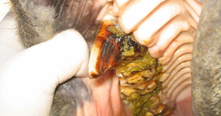
Figure 1. Large focal overgrowths of the 106 cheek tooth.
Dental overgrowths are commonly encountered in equine practice for a variety of reasons.

Perhaps the most commonly observed overgrowth is attributable to the anisognathic conformation of the horse’s head, whereby the maxilla is wider than the mandible, the effects of which can be exacerbated by feeding high levels of processed food. This contributes to “overgrowths” of sharp enamel points on the buccal aspects of the maxillary and lingual aspects of the mandibular arcades.
This anatomical predisposition is compounded by the effects of domestication and the reduced mastication and altered chewing cycle of concentrate diets (Carmalt and Allen, 2006; Carmalt and Carmalt, 2007).
Equine teeth continue to erupt at a fairly constant rate (2mm/year to 3mm/year) and, providing the horse is fed a predominately fibrous diet, wear (attrition) from the herbivorous diet tends to keep pace with eruption rates. Discrepancies in length and position of the mandible and maxilla can also cause the frequently encountered problem of rostral (06) and caudal (11) focal overgrowths (Figure 1).

Overgrowths generally occur when there is a lack of opposing occlusion (Figure 2), such as:
A single unopposed tooth out of occlusion will become progressively overgrown through lack of attrition and greatly increased eruption of an unopposed tooth. This type of scenario can often result in a vicious cycle of a disrupted masticatory pattern and subsequent generalised overgrowth patterns, such as “wave complexes” or “shear mouth” (Dixon et al, 1999; Figure 3).
Early recognition and appropriate treatment of dental overgrowths is, therefore, paramount for equine welfare, as mucosal injuries are associated with sharp enamel points and dental overgrowths in the horse (Salem et al, 2015).
The principles in treating dental overgrowths are the same for all; safe reduction of the overgrowth, to take the damaged or worn surface of the opposite arcade out of occlusion, prevent soft tissue damage, and restricted rostrocaudal and lateral jaw movement.

The advent of motorised dental equipment has enhanced the practitioner’s ability to reduce overgrowths efficiently and accurately, although research has highlighted this is not without iatrogenic risk to pulp tissues.
It is worth mentioning, in the UK only veterinary surgeons and “qualified” equine dental technicians (EDTs) are permitted to use motorised dental equipment and “horses should be sedated unless it is deemed safe to undertake any proposed procedure without sedation, with full informed consent of the owner” (BEVA, 2015).
Reduction of overgrowths should be carried out under continual visual assessment using an appropriate light source and dental mirror/oral endoscope, with the horse suitably sedated and restrained. Many varieties of motorised dental equipment are available and, given the potential for iatrogenic damage, individuals should have received appropriate training to ensure an efficient and safe technique is utilised for reduction of dental overgrowths.
A great potential exists for pulpar exposure or damage to pulps that have not been exposed from over-zealous use of motorised dental equipment. Any energy not used in the actual grinding process will be transformed to heat, resulting in thermal damage (Wilson and Walsh, 2005). The amount of heat transformed will depend on the type of equipment – including speed and torque, and ability to water cool – in addition to rasping time, environmental factors and distance of pulp tissue from the dental tissue (O’Leary et al, 2013).
Corrective procedures should maintain or attempt to establish correct cheek teeth (CT)occlusion and symmetry, but one should be aware some major disorders cannot, and should not, be fully corrected at a single corrective dentistry session without compromising the long-term dental life of the horse.

The approach to overgrowth reduction should be one of a staged attenuation of 3mm to 4mm at a time, especially if the overgrowth has occurred over a period of months/years. There should be constant assessment (visually with a dental mirror or oral endoscope) during the procedure to prevent pulp exposure and maintain occlusal angles.
Particular care must be taken with geriatric patients, although the perceived risk in reducing overgrowths can be less than in younger patients. Studies have shown subocclusal secondary dentine depth can be even more variable (and less thick) in this cohort (Marshall et al, 2012).
Overgrown teeth in geriatrics tend to be further complicated by the lack of reserve crown, which can lead to instability in the alveolus and loss or displacement of these teeth, in addition to concurrent concerns with sedation and restraint in these patients (Figure 4).
The majority of cheek teeth (07 to 11) have five pulp chambers each, although the rostral and caudal cheek teeth (06 and 11) have additional pulp horns. This creates potential risks and complications when the rostral aspect of the 06s are “profiled” aggressively (previously termed “bit seating”), a procedure that has no scientific basis. The depth of subocclusal dentine is particularly variable at this location, so the validity of aggressive profiling of the 06s should be questioned (Figure 5).

Pulp exposure and thermal damage may take time to become clinically apparent, and as practitioners, we have moved away from the procedure of bit seating as knowledge and understanding of equine dentistry has progressed. Consequently, reduction of these overgrown teeth to the level of adjacent normal-height teeth has been reported to cause occlusal pulp exposure in 58% of teeth, in addition to possible thermal damage (Marshall et al, 2012).
Therefore, equine CT overgrowths should be gradually reduced in stages, by a few millimetres per treatment and over a prolonged period of time. Marshall et al (2012) showed if overgrowths were reduced to the level of the adjacent tooth, 58% of these procedures would result in pulp horn exposure.
The advent of motorised dental equipment has potentially exacerbated the additional risks of thermal damage during overgrowth reduction, as evidenced by a study from the Edinburgh group (O’Leary et al, 2013), which found prolonged rasping with three different types of motorised dental equipment increases the likelihood of heating a pulp above a critical damage. Almost 20% of teeth that were reduced for 30 seconds exceeded critical temperature thresholds for thermal damage. However, increased dentinal thickness, reduced duration of use and water cooling had protective roles in reducing thermal damage.
The take home message is to perform any reduction carefully, accurately and conservatively – you cannot put dental tissue back on.