31 Aug 2015
Moses Brennan
Job Title
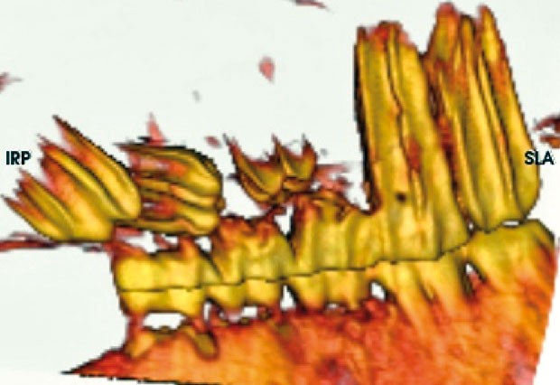
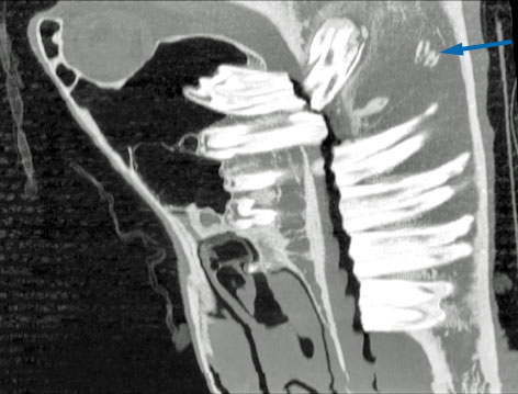
Figure 8. 3D volume-rendered reconstruction of an 11-year-old mare with a large mass on the lateral aspect of the right mandible. Histopathological analysis of biopsies confirmed the presence of an aneurysmal bone cyst.
CT of the horse’s head is widely available in equine practice and its use in providing accurate images of dental anatomy and pathology is rapidly becoming the gold standard for investigation of dental disease.

A system that allows scanning of the standing, sedated equine patient was first developed by the late Alastair Nelson at the Rainbow Equine Hospital, North Yorkshire in 2003. The system uses a platform that floats on compressed air skates and has been installed in eight UK hospitals with various modifications (Figure 1). This system allows the safe and speedy acquisition of CT images of the head and proximal cervical regions.
The great advantage of CT over conventional imaging techniques is it allows the images to be viewed in three planes – frontal, sagittal and longitudinal – without the superimposition of the complex anatomical structures of the equine head. Three-dimensional reconstructions of the data acquired also allows for great flexibility in image manipulation and is a very useful technique for providing additional information for pre-surgical planning of the most challenging cases.
CT has been shown to have a high sensitivity and specificity in the diagnosis of dental disease when compared to radiography1.

As in most mammals, the equine tooth is composed of enamel, cementum, dentine and dental pulp and is supported by the periodontium (periodontal ligament, alveolar bone and gum)2. The unit of measurement of a material’s density on CT is the Hounsfield unit, and each material has a different measurement, which doesn’t vary with age. CT allows manipulation of the window levels, allowing optimal visualisation of each structure with a different density (Figure 2)
Due to the hypsodont nature of the equine tooth, with its continuous eruption, there is a constant change in the morphology of the tooth over time. Recently, the detailed CT anatomy of the equine tooth has been described3. This research showed the number of interpulpar communications between pulp horns and the pulpar volume of each tooth decreased with increasing age. The study also showed the interpulpar communications of the maxillary teeth were of greater complexity and variety than the mandibular teeth.

Cemental hypoplasia most commonly affects the apical aspect of the infundibulum and is a common finding in maxillary cheek teeth. Some authors consider this to be a normal finding4. More recent research identified only 10% of infundibulae to have no lesions visible either on the occlusal surface or on CT examination and concluded some infundibular changes associated with age could be considered normal5 (Figure 3).
Dental caries has been defined as a progressive acidic demineralisation of the inorganic matrix of dental tissues secondary to bacterial fermentation of impacted carbohydrate substrate and subsequent organic matrix loss6. Once infundibular hypoplasia becomes exposed at the occlusal surface, caries is invariably present and so the combined term infundibular hypoplasia/caries has been used7.

A grading system for infundibular caries has been established, based on the structures involved and the severity of the lesion:
CT allows a more thorough evaluation of the extent of infundibular lesions, especially if treatment by filing of these lesions is to be considered.
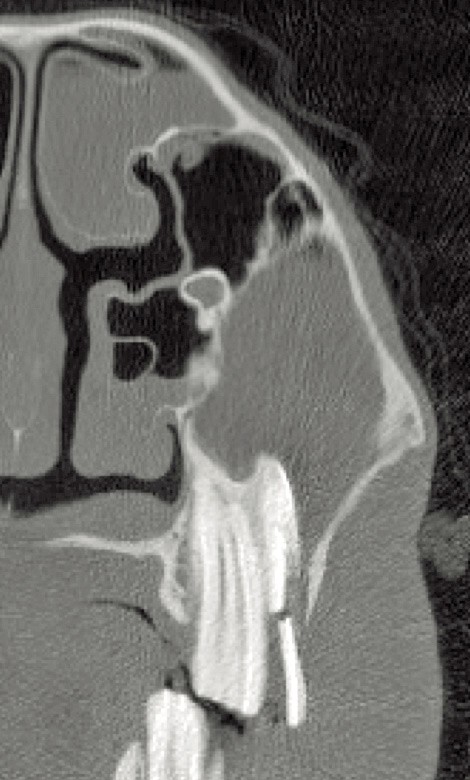
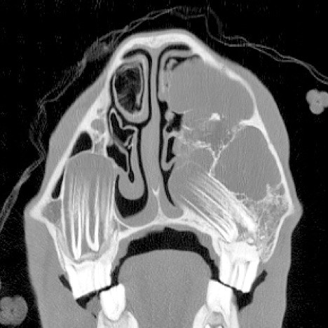
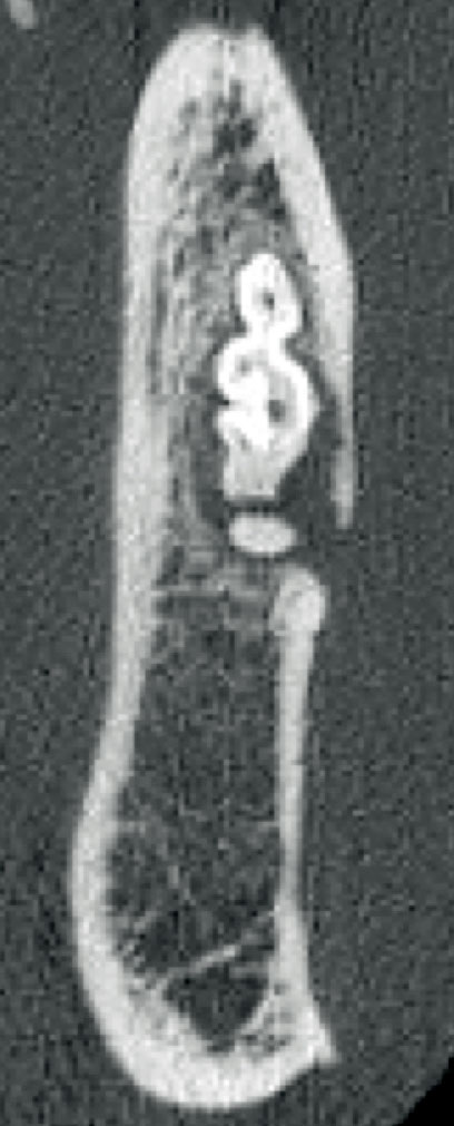
Idiopathic slab fractures of the maxillary 09 are the most common type of dental fracture in equine teeth – accounting for approximately 80%2. The exact reason these fractures occur remains uncertain; however, in some instances, they can enter the pulp horns and lead to secondary sinusitis. CT examination can allow accurate identification of pulpal involvement and can predict which teeth should be removed to reduce the risk of further complications (Figure 5).
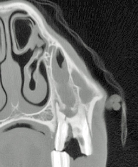
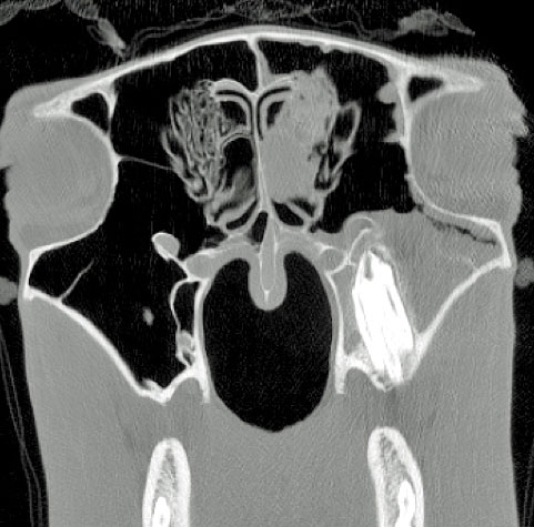
The remainder of idiopathic dental fractures are midline sagittal fractures that originate as infundibular caries. These, again, can occur with or without an associated secondary sinusitis (Figure 6).
Although very common and of great clinical significance, the exact aetiopathogenesis of primary apical infections is poorly understood. It is thought an acute pulpitis resulting from bacterial infection of the pulp chamber is the cause. Several mechanisms for the entry of the bacteria into the pulp chamber have been proposed; however, the most likely mechanism involves the seeding of bacteria into areas of pulpal inflammation9. In a case series, patent infundibulum resulting in apical infections of check teeth was described10. Secondary apical infections can also occur due to polydontia, dental displacement, dysplasia and fractures of the teeth or bone.
Signs of apical infection that can be identified with CT include:
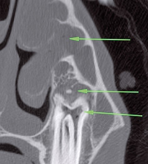
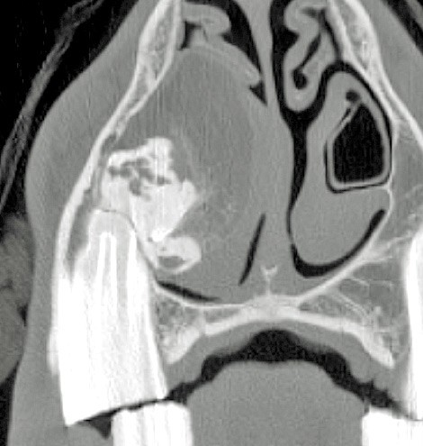
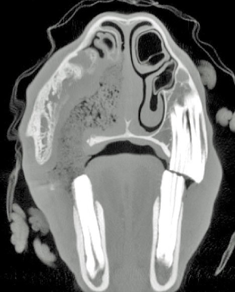
Gas bubbles within the bulging root area, or fragmentation of the root in combination with swelling of the adjacent sinus lining have been described as the most important CT features of dental decay11 (Figure 7).
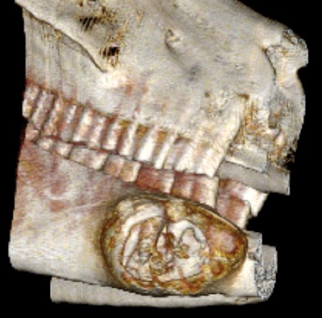
Odontogenic tumours are rare in general, although they seem to be more frequent in horses than in other species2. CT is useful when trying to plan surgical treatment of such lesions (Figures 8 and 9).
Supernumerary and displaced cheek teeth can occur in very challenging areas to access surgically. The ability to accurately plan surgical access with the benefit of CT is essential when dealing with these challenging cases (Figures 10 and 11).
CT can be an excellent tool for investigating troublesome discharging sinus tracts post-extraction. It is not uncommon to find enamel root fragments of CT images of horses that have previously had teeth removed via repulsion. In some instances, these root fragments may be asymptomatic (Figure 12).
CT can also be useful when investigating orosinal or oronasal fistula as a complication of cheek tooth extraction (Figure 13).
CT provides unsurpassed detail when imaging the equine dental patient. With its use becoming more widespread, our understanding of normal dental anatomy and pathology is increasing, allowing the development of less invasive and more accurately targeted treatment techniques.
CT is widely available for imaging the equine head. When imaging complex anatomical areas CT provides a far superior level of information regarding normal anatomy and subtle pathology that can be easily missed with conventional imaging techniques. CT can also be extremely useful when dealing with more complicated dental disease in the equine patient.