3 Sept 2024
David Charles discusses the considerations for farm vets facing emergency call outs for this condition.
David Charles
Job Title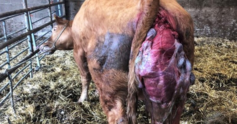
Image: David Charles
Bovine uterine prolapses are an uncommon, but important, emergency call attended by cattle vets.
While the basic technique for replacement of the prolapsed uterus has not fundamentally changed in the past 10 to 20 years, much has been learned regarding causation, success rates and appropriate aftercare.
This article will review the causes of bovine uterine prolapses, offer guidance to vets regarding replacement, and discuss appropriate aftercare. Lastly, we will review the literature to aid vets in providing an evidence-based prognosis to clients.
Numerous potential causes of uterine prolapse in cattle are reported in the literature, and it has a reported incidence rate of 0.002% to 1% of calvings (Correa, Erb and Scarlett, 1992; Miesner and Anderson, 2008; Martin et al, 2023).
A Carluccio et al (2020) study of 33,450 calving cows (5,700 beef and 27,750 dairy cattle) reported a greater prevalence in beef cattle (1%) compared with dairy cattle (0.6%).
Uterine prolapse has been demonstrated to be associated with dystocia, hypocalcaemia and, in some studies, age, although this could be due to its correlation with hypocalcaemia (DeGaris and Lean, 2008; Valldecabres, Pires and Silva-del-Río, 2019). It is believed that (rarer) delayed uterine prolapse tends to be caused by failure of closure of the cervix.
Hypocalcaemia can cause a reduction in uterine tone and weakened myometrial contractility. Risco et al (1984) took coccygeal blood samples from 106 cows postpartum (53 with uterine prolapse and 53 without uterine prolapse). They found cows with uterine prolapse had a significantly lower serum calcium concentration (mean serum calcium concentration 1.52mmol +/- 0.06) than those without.
As with most procedures in farm animal practice, a client will need to wait a period of time for the attending vet to reach the farm. During this time the author recommends clients advise farmers of the following:
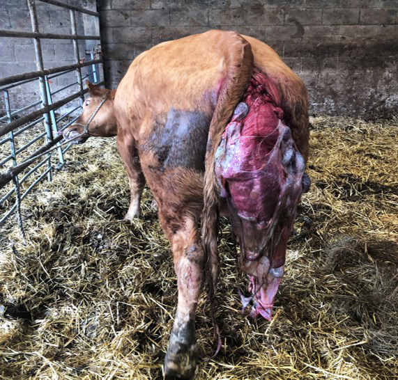
If the cow is standing, careful consideration of its restraint is required. If the cow can be safely, and calmly, moved to a crush or a calving gate, this is recommended (Figure 1). Damp sheets can be used to “wrap” the prolapse to protect it before walking the cow to the crush. Sometimes, it is also possible to lift the prolapse and follow closely behind the cow to reduce the risk of trauma from the legs, feet or gravity during movement.
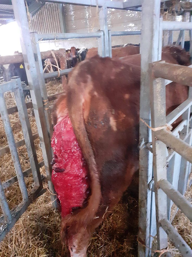
Sometimes a crush, calving gate or lock-in head yolk is not available at the cow’s location. In these scenarios it may be that haltering the cow to a secure fence or other immovable object is the only option. Vet and farmer safety must be protected in these scenarios, and standing sedation of the patient may be necessary. The cow in Figure 2 was one such example, whereby the cow was in a loose calving box and the farmer didn’t have a crush or other equipment. Therefore, a halter was applied, and after the photograph was taken IV sedation with 70mg of xylazine hydrochloride was administered to provide adequate standing sedation (Lin, 2022) for safe replacement of the prolapse.
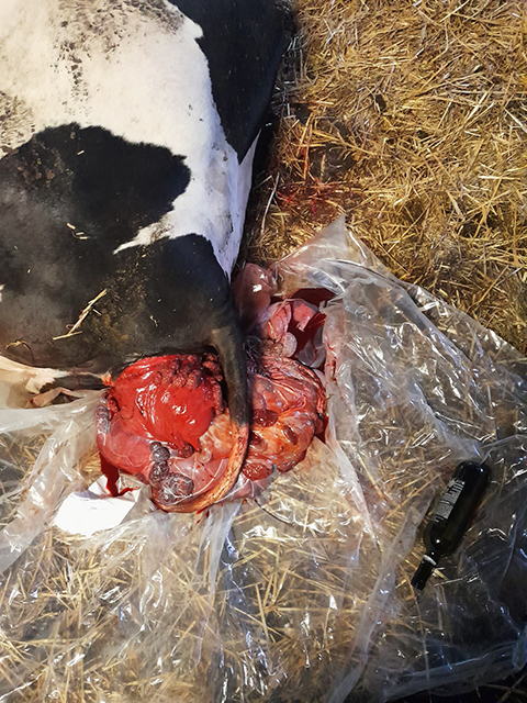
If the cow is recumbent (Figure 3), it needs to be placed into a stable sternal recumbency, and the legs positioned in the “New Zealand” position (colloquially referred to as “frog-legged” or “spatchcock chicken” positioning). This provides optimal positioning for replacement of the uterus, tilting the pelvis downward by approximately 30˚ (Weaver et al, 2018).
This can require significant physical exertion and sufficient numbers of people should be sought to achieve this; it is also worth noting that if the cow is in sternal recumbency for the procedure, an assistant wearing appropriate PPE can stand/sit astride the cow and lift the uterus from above to aid repositioning.
Some cattle with uterine prolapses present in hypovolaemic shock, due to significant internal bleeding, organ prolapse or entrapment of abdominal viscera (Potter, 2008). IV fluid therapy may be required in these cases (Roussel, 1990), and careful consideration should be given to the prognosis given to the farmer. In some cases (for example, if a significant uterine artery bleed has already occurred prior to arrival) it will be important to discuss the cost versus prognosis analysis with the farmer.
The aim is to safely restore the uterus to the everted position. The below steps detail an approach to the replacement of the bovine uterine prolapse. Variations on this approach are described in the literature, and practitioners will have their own “tips and tricks” that aid them in replacement. The following is the favoured approach by the author based on clinical experience and review of the evidence base.
Debate continues among farm vets as to whether to place a perivulval suture after the replacement of a prolapsed uterus. Therefore, suturing is not universally practised. However, geographical variation can be seen. In a Norwegian study (Martin et al, 2023), 98% of respondents placed a perivulval suture. In the author’s experience, use of vulval suture by UK practitioners is more variable.
If a suture is to be placed, use of a sharp sterile Buhner needle with 6mm nylon thread or 7mm Buhner tape can reduce the risk to the tissue if repeat prolapse occurs. Additionally, stents can be added in, if desired, to reduce the risk of tearing. Practitioners should ensure the cow can still urinate once the suture is tied.
Oxytocin (10IU to 40IU) should be administered to promote involution of the uterus, and aid passage of any placenta left in situ. Analgesia (NSAIDs) are strongly recommended for three to five days.
As environmental contamination almost always occurs, the author believes antibiotics can be justified. However, a broad-spectrum, category D antibiotic should be prescribed. The author favours an initial course of three days of amoxicillin or penicillin. For beef cattle, where handling may be harder, long-acting preparations of procaine penicillin G are available, with a single dose of 20mg/kg providing 72 hours of antimicrobial activity.
Calcium should be provided, owing to the close association between uterine prolapse and hypocalcaemia. For rapid elevation of plasma calcium concentration, IV calcium borogluconate 40% can be administered, but its effect is short-lived (Blanc et al, 2014).
Cows with uterine prolapses are often dehydrated, and pump-drenching 20L of warm water made up with a product containing 500g of calcium propionate can provide rehydration, while also providing a more sustained increase in plasma calcium concentrations compared with IV calcium borogluconate or oral calcium chloride products (Goff and Horst, 1993; Blanc et al, 2014). Oral calcium propionate given shortly after calving has also been demonstrated to reduce the incidence of metritis (Stokes and Goff, 2001).
If successful replacement of the uterus can be achieved, prognosis is favourable. Vets should be aware of an occasional risk of death after, or during, replacement due to rupture of the uterine artery (Table 1). Vaginal prolapse prepartum or significant uterine oedema and/or trauma have been demonstrated to reduce survival rate (Murphy and Dobson, 2002; Martin et al, 2023).
| Table 1. Survival rates and conception rates in subsequent lactation reported in literature for cows undergoing replacement of a uterine prolapse | ||
|---|---|---|
| Author | Survival rate (percentage) | Conception rate in subsequent lactation (percentage) |
| Martin et al, 2023 | 72 at 30 days | N/A |
| Carluccio et al, 2020 | 81.9 | 84.7 |
| Martin et al, 2023 | 72 at 30 days | N/A |
| Ishii et al, 2010 | 64.7 | 80.6 |
| Murphy and Dobson, 2002 | 80 | 80.5 |
| Gardner et al, 1990 | 72.4 | 81.25 |
Interestingly, one study (Martin et al, 2023) reported the time taken to replace the uterus dramatically affected survival rates 30 days post-procedure. If it took more than 20 minutes to replace the prolapsed uterus, the hazard of death was three times greater.
Repeat uterine prolapse after subsequent calvings appears to be rare. Both Jubb (1990) and Anderson (2012) report below 1.5% of their study populations (158 in total) either had suffered from uterine prolapse in previous lactations or re-prolapsed in subsequent years.
Uterine prolapse remains an uncommon, but important, obstetrical condition of cattle. If successful replacement can be achieved, survival rates are favourable. Having a methodical approach, and being familiar with the technique, is important for the vet doing any ambulatory farm work.
As new studies have shown the impact of replacing the uterus within 20 minutes of starting, vets should ensure they are appropriately prepared prior to commencing attempted replacement.