Register

Login

Vet Times logo
+
  • View all news
  • Vets news
  • Vet Nursing news
  • Business news
  • + More
    • Videos
    • Podcasts
    • Crossword
  • View all clinical
  • Small animal
  • Livestock
  • Equine
  • Exotics
  • All Jobs
  • Your ideal job
  • Post a job
  • Career Advice
  • Students
About
Contact Us
For Advertisers
NewsClinicalJobs
Vet Times logo

Vets

All Vets newsSmall animalLivestockEquineExoticWork and well-beingOpinion

Vet Nursing

All Vet Nursing newsSmall animalLivestockEquineExoticWork and well-beingOpinion

Business

All Business newsHuman resourcesBig 6SustainabilityFinanceDigitalPractice profilesPractice developments

+ More

VideosPodcastsDigital EditionCrossword

The latest veterinary news, delivered straight to your inbox.

Choose which topics you want to hear about and how often.

Vet Times logo 2

About

The team

Advertise with us

Recruitment

Contact us

Vet Times logo 2

Vets

All Vets news

Small animal

Livestock

Equine

Exotic

Work and well-being

Opinion

Vet Nursing

All Vet Nursing news

Small animal

Livestock

Equine

Exotic

Work and well-being

Opinion

Business

All Business news

Human resources

Big 6

Sustainability

Finance

Digital

Practice profiles

Practice developments

Clinical

All Clinical content

Small animal

Livestock

Equine

Exotics

Jobs

All Jobs content

All Jobs

Your ideal job

Post a job

Career Advice

Students

More

All More content

Videos

Podcasts

Digital Edition

Crossword


Terms and conditions

Complaints policy

Cookie policy

Privacy policy

fb-iconinsta-iconlinkedin-icontwitter-iconyoutube-icon

© Veterinary Business Development Ltd 2025

IPSO_regulated

30 Oct 2017

Farm animal pathology and disease surveillance: a new era

Chris Palgrave, Pernille Jorgensen and David Harwood discuss the inception of a network of veterinary investigation centres, plus its evolution of aims and objectives.

Chris Palgrave, David Harwood, Pernille Jorgensen

Job Title


Farm animal pathology and disease surveillance: a new era

Postmortem examination (PME) of livestock, and the information it generates, is an important part of a farm animal health programme. This article explores how a national network of veterinary investigation centres (VICs) was set up, and how the service has evolved as its aims changed over time.

Livestock keepers and their vets are the obvious beneficiaries, but underpinning this is a continuing need to ensure the UK has a robust system in place to protect the consumer (and the taxpayer) from any future animal-related public health risk, such as BSE. Each PME is, in essence, a fragment of information that when combined with other such fragments is able to build up a valuable overview of what is a dynamic and ever-changing UK disease status.

Deadstock at a fallen stock collection centre – are we missing anything?

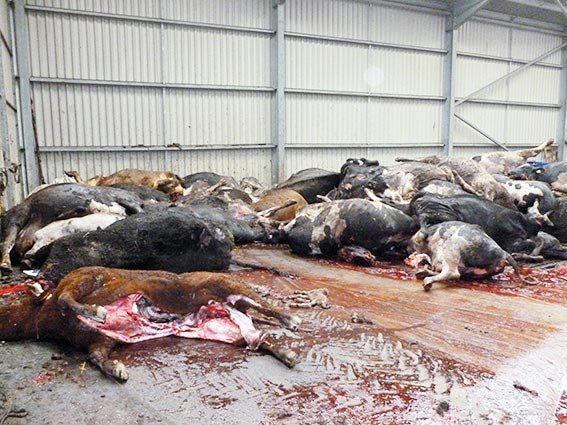
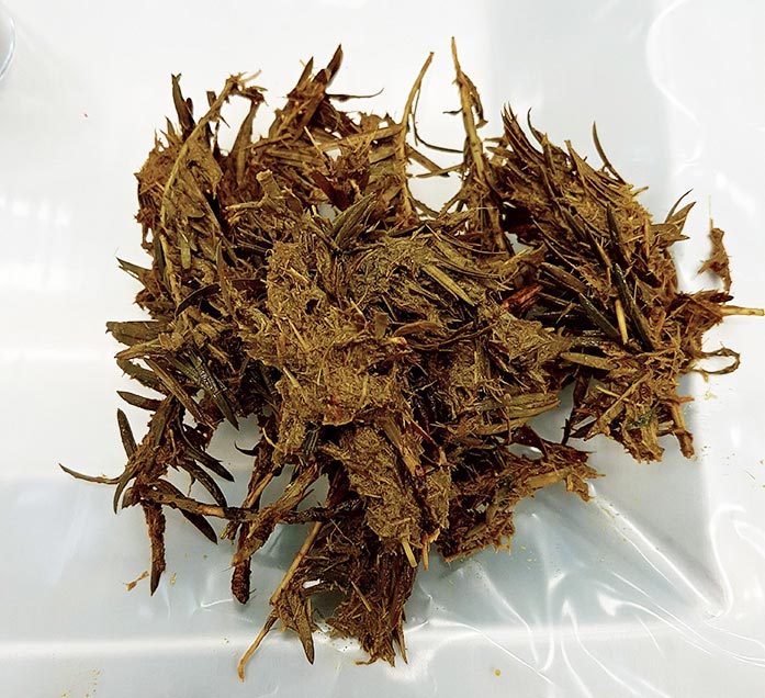
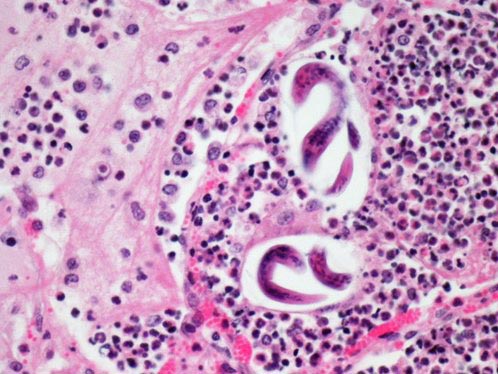
The image to the right shows a typical day’s mortality collected in a busy fallen stock centre. Are we in danger of missing something if we routinely ignore such losses when they begin to escalate on any particular holding?

Background

In 1894, in response to a severe swine fever outbreak, the first national veterinary laboratory was established in a small basement room at Whitehall. This was the forerunner to the Central Veterinary Laboratory, which has been on its site in Weybridge since 1917.

Later, in 1922, the first network of veterinary laboratories was developed, operating in existing agricultural colleges. This gradually evolved into a national network of VICs established across England and Wales, providing both an important diagnostic service to vets and farmers, and a valuable source of surveillance data to monitor endemic disease, as well as detect new and emerging diseases.

Over decades, this network grew to 24 VICs and played a pivotal role in identification and control of not only important farm livestock diseases, but also newly recognised conditions.

An adult bull ready for postmortem examination.

This network remained static until a series of reviews – Rayner in 1984, followed by Dalton in 1990 and, most recently, Surveillance 2014 – took place. The number of centres (now referred to as APHA VICs) was reduced significantly to six – Bury St Edmunds, Carmarthen, Penrith, Shrewsbury, Starcross and Thirsk, in addition to APHA Lasswade, which focuses on avian PMEs. This potentially left significant areas of England and Wales without a convenient local VIC, posing a challenge for farm animal diagnostics and effective disease surveillance.

Initially, an interim government-funded transportation service was put in place, collecting carcases from farms and delivering them to the nearest APHA VIC. However, since September 2014, a number of designated “third-party providers of postmortem facilities”, operating under contract to the APHA, have been providing cover to areas no longer served directly by an APHA VIC. These include the University of Surrey (Guildford), RVC (North Mymms), University of Bristol (Langford) and Wales Veterinary Science Centre (Aberystwyth).

This has been augmented by a free carcase collection service that, since January 2017, has provided coverage for all farms outside a one-hour drive time to a PME site.

Severe fibrinosuppurative (“bread and butter”) pericarditis in a cow with traumatic reticulopericarditis following ingestion of a piece of wire from a tyre.

All APHA VICs, together with third-party providers, contribute to the surveillance-gathering programme, which also includes the network of SAC Consulting Disease Surveillance Centres in Scotland. In addition to the official centres, a number of independent PME providers exist, including the University of Liverpool Institute of Veterinary Science, Farm Post Mortems (north-east England) and Veterinary Investigation Services (Gloucestershire), which contribute to the network through discussions and informal sharing of surveillance information.

Data analysis and results sharing

The national scanning surveillance programme is underpinned by powerful analysis of detailed data gathered by all members of the official network. At the heart of this are the Veterinary Investigation Diagnostic Analysis (VIDA) criteria, in which every diagnosis recorded is allocated a code and kept under constant review to assess trends or changes that may be important.

In addition to many diagnoses successfully reached, particular attention is paid to two additional categories – “diagnosis not reached” and “diagnosis not listed” – as it is potentially in these categories where something “new and unusual” may be identified.

A composite monthly surveillance summary appears in Veterinary Record and the analysis of VIDA data is shared through the species quarterly emerging threat reports1. Newsletters are also produced by the various providers as a means of communicating information.

However, the way that VIDA and other surveillance data is provided is rapidly evolving; for small ruminants and cattle, surveillance data can now be viewed in real time via a web-based disease surveillance dashboard2. Similar ones will be made available for other species.

Caudal vena cava abscessation and thrombosis in an adult Holstein cow with rumenitis.
evere icterus as a result of copper poisoning in a calf.
Severe icterus as a result of copper poisoning in a calf.
Yew leaves and twigs removed from the rumen of a cow that died suddenly following trimming of a hedge.
Cortical necrosis and haemorrhage in the kidney from a lamb that died suddenly due to clostridial enterotoxaemia (“pulpy kidney”).
A placenta from an aborted lamb – severe intercotyledonary necrosuppurative placentitis due to Chlamydophila abortus (enzootic abortion of ewes).
The oral cavity/head of an aborted lamb with severe palatoschisis (cleft palate) and cheiloschisis (cleft lip).

Problem of selection bias

Chicken – distended loops of small intestine with black and white “salt and pepper” foci (left) containing watery to mucoid orange foamy contents (right) due to coccidial enteritis.

One insurmountable problem is selection bias. The typical sequence of events is as follows:

  • A livestock keeper will contact the veterinary practice to discuss a dead, sick or “problem” animal or bird; this may be a single incident or part of an ongoing issue.
  • In some cases, a vet may be familiar with the farm, make routine visits and be aware of the problem at hand. In other cases, it may have been months (or years) since the last visit and the telephone call comes out of the blue.
  • In most cases, it is the vet who suggests a PME may be of value, although livestock keepers are increasingly initiating this conversation.
  • Vets may elect to carry out a PME on the farm or, in some cases, at a fallen stock site. The latter option can be beneficial (and safer), as lifting equipment for manipulating heavy carcases, help on hand (if needed) and easy disposal of the carcase/viscera is usually available. In some cases, a designated area away from the main thoroughfare exists where carcases can be held and examined.
  • While many farm animal vets have considerable skill and experience at performing PMEs, a point may come where they would like to refer a carcase for examination at their closest PME facility.

Why might a PME help?

Many scenarios exist where a PME can be useful:

  • A genuine “sudden death”, where an urgent need exists to find out why the animal died, ensuring anthrax and/or other notifiable disease is considered at the outset.
  • Urinary bladder of an adult cow with multiple haemorrhage polypoid (finger-like) mucosal masses due to chronic bracken toxicity (bovine enzootic haematuria).
    Urinary bladder of an adult cow with multiple haemorrhage polypoid (finger-like) mucosal masses due to chronic bracken toxicity (bovine enzootic haematuria).

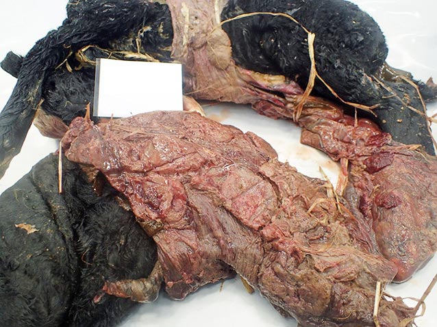
    An animal dying during a disease incident, such as pneumonia in calves, pyrexia and weight loss in an adult cow or unexpected periparturient ewe losses. It is important to ensure any carcase submitted is typical of the incident, and preferably one that has received little, if any, treatment.

  • Investigating causes of abortion and stillbirth.
  • Follow-up to a clinical or surgical case a vet has been dealing with.
  • Possible product failure or adverse reaction to a product (often supported by a pharmaceutical company). It is important to ensure a Suspect Adverse Reaction Reporting Scheme (SARRS) report is made to the VMD.
  • Suspect toxicity incidents.
  • Sacrificial PME in a problem, such as ill-thrift in lambs and diarrhoea in pigs. In these cases, always ensure, where possible, a full range of blood samples is taken prior to euthanasia.
  • Welfare investigation (forensic) – often instigated by a local authority, the RSPCA or the APHA.

I would like to send a carcase for PME, what should I do?

To send a carcase for PME, firstly, it is important to identify the appropriate PME provider by entering the postcode of the farm into the APHA postcode finder3. This will also inform you of whether the holding is eligible for the free carcase collection service. Farms within a one-hour radius of the PME provider must arrange delivery of the carcase. Those more than one hour away are eligible for free collection.

Secondly, as the cost of the PME and any additional testing is heavily subsidised by the APHA, you must telephone the provider to make sure the carcase is eligible (see further on). If appropriate, the PME provider will instruct the haulier to collect the carcase.

The thickened small intestine from a sheep. Left: crypts separated by sheets of macrophages in the lamina propria (haemotoxylin and eosin). Right: macrophages contain vast numbers of intracellular acid-fast (Ziehl-Neelsen, bright pink) bacilli consistent with Mycobacterium avium paratuberculosis (Johne’s disease).

The triage process assesses the time that has elapsed since death, the number of animals from a holding presenting with the same condition, the chronicity of the condition, treatment and the potential for notifiable disease. Some PME providers may still accept carcases that fail the triage process, but these are not eligible for the APHA subsidy and the cost of any additional testing may not be included in the PME price.

How to maximise diagnosis chances – time is of the essence

To maximise the chance of getting a diagnosis:

  • Encourage your clients to telephone as soon as possible. The dead animal may not get priority over other important parts of the daily routine, but it is essential they set the ball rolling as soon as possible to expedite delivery to the postmortem provider and maximise the diagnostic quality of the carcase.
  • In the case of abortions/stillbirth, it is vital to include the placenta, if at all possible.
  • Detailed information is required for the triage process; therefore, instruct any practice staff who may be taking telephone calls from farmers to ask all the pertinent questions:
    • A lung from an adult goat with pneumonia: two coiled nematode larvae (Muellerius capillaris) are surrounded by degenerate neutrophils and macrophages.
      A lung from an adult goat with pneumonia: two coiled nematode larvae (Muellerius capillaris) are surrounded by degenerate neutrophils and macrophages.

      the owner’s name, address, postcode, telephone number and, if possible, County Parish Herd Holding number

    • species, breed and age of animal(s) affected
    • time of death and number affected/dead
    • number of animals in the herd/flock and in the same group as the affected animal(s)
    • a brief history, including signs and duration
    • treatment given and relevant management procedures, vaccination history and nutrition
    • any suspicion of notifiable disease
    • contact name of the veterinary surgeon in the practice
  • Use the postcode finder and the first part of the farm’s postcode to identify the nearest PME provider to determine if it is eligible for the free collection service.
  • Telephone the relevant PME provider as soon as possible. It will use the information provided by the farmer to triage the case and determine whether he or she is eligible for an APHA-subsided PME. The provider will also instruct the haulier to collect the carcase, if appropriate.
  • If the farm is not eligible for free carcase collection, ensure the postmortem facility location is known to anyone delivering his or her own carcase.

University of Surrey Veterinary Pathology Centre

The main postmortem room at the University of Surrey Veterinary Pathology Services Centre.

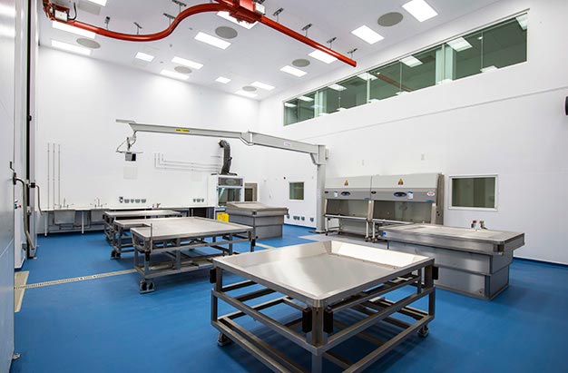
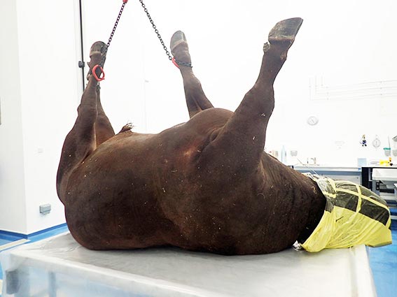
The University of Surrey is the designated third-party PME provider for south-east England, and has a modern, purpose-built Veterinary Pathology Centre that is part ofthe university’s school of veterinary medicine in Guildford4.

It is staffed by a team of technical and administrative staff, veterinary investigation officers and board-certified veterinary pathologists. On completion of the PME,APHA laboratories and third-party providers aim to provide an initial verbal and/or written report the same day or early the following day. This may be followed by the results of additional testing as they are received.

Once all further testing is complete, a final report is produced summarising the case, and provides the diagnosis (where possible) and additional comments.

While, despite all our best efforts, it is not always possible to reach a diagnosis in every case, we do achieve an 85% diagnostic rate. Where a disease is not diagnosed, this may warrant further investigation and support to take cases forward is available.

Carrying out your own PME?

Veterinary staff at any of these designated locations are available for case discussion and sampling requirements. The APHA’s guide on sample and test selection5 provides guidance on the samples required and tests used for common disease presentations for both livestock and wildlife6.

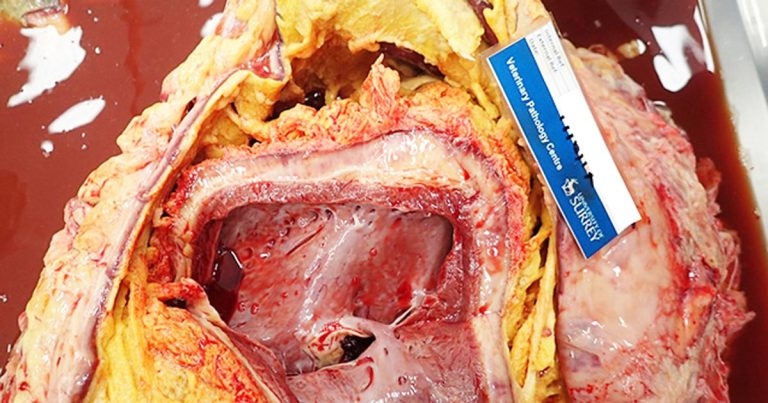
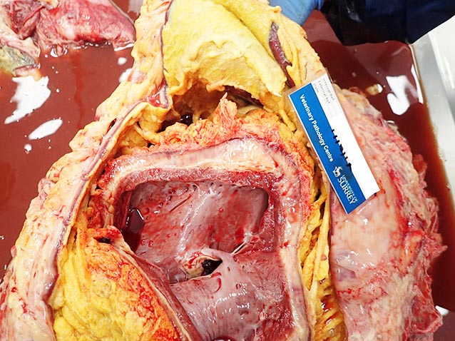
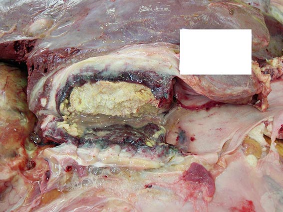
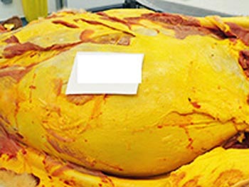
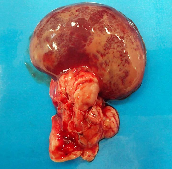
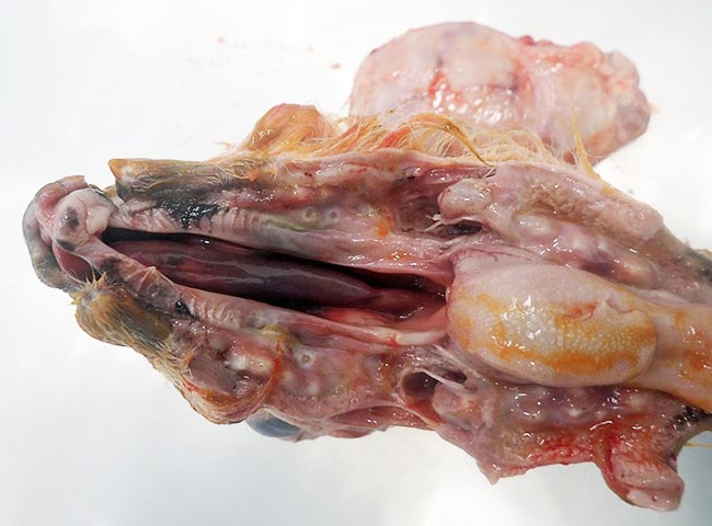
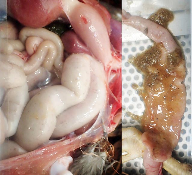
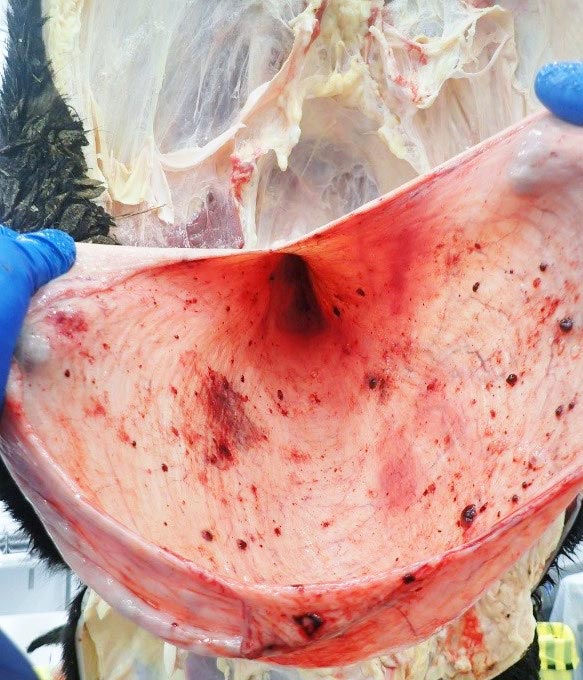
Good images of postmortem lesions can be particularly useful. If you encounter anything that looks unusual and/or interesting, do not keep it to yourself – share it with colleagues and the wider veterinary surveillance community.

Summary

New and emerging problems have to start somewhere and it could easily be with your and/or your client.