20 Dec 2018
Axiom Veterinary Laboratories assesses a series of case studies of infections on farms, including viral respiratory and bacterial respiratory infections.
James Barnett
Job Title
Presented are selected cases from the ruminant diagnostic caseload of Axiom Veterinary Laboratories.
Axiom provides a farm animal diagnostics service to more than 300 farm and mixed practices across the UK, and receives both clinical and pathological specimens as part of its caseload. The company is grateful to clients for the cases presented in this article.
Several cases of lungworm, Dictyocaulus viviparus, were diagnosed on Baermanns, including one four-year-old suckler cow potentially under-dosed with doramectin and, in a second herd, two coughing, scouring Friesian heifers, treated four weeks earlier with levamisole. False negatives will occur with Baermann testing if faeces samples are not refrigerated and submitted to the laboratory quickly, preferably on ice packs.
Seroconversion to lungworm was seen in ill, thrifty, coughing 14-month-old to 18-month-old beef cattle and, in a second herd, in a similarly affected three-year-old cow. In a third herd, milk drop was the main presenting sign. Following challenge, naïve animals seroconvert after approximately four to six weeks and titres remain elevated for four to five months. Therefore, positive results usually indicate exposure in the current grazing season.
Lungworm also was diagnosed on histopathology of the lung from a one-year-old beef animal. The histological appearance was of a marked interstitial type pneumonia and bronchitis/alveolitis, and frequent intralesional helminth larvae, were detected – with some adult helminth parasites consistent with D vivparus in larger bronchioles.
PCR has largely replaced indirect fluorescent antibody test for the detection of viral antigen on nasopharyngeal swabs and in bronchoalveolar lavage and tissue samples, but animals sampled need to be in the acute stage of disease to maximise the chances of detecting virus.
Infectious bovine rhinotracheitis (IBR) infection was diagnosed by PCR on pooled swabs from 18-month-old cattle bought-in in July and vaccinated with an IBR marker vaccine. Clinical signs included ocular signs and pyrexia. Similarly, IBR was detected in 2 of 50 recently bought-in beef finisher bulls vaccinated with live marker vaccine shortly after arrival, but developed upper respiratory tract signs and pyrexia. The bulls were also bovine viral diarrhoea viraemic. In a third case, IBR was confirmed by PCR in a pyrexic, tachypnoeic heifer with serous nasal discharge.
RSV was diagnosed by PCR on the lung of a four-month-old calf with cranioventral pulmonary consolidation on postmortem and in a pre-weaned calf with pulmonary consolidation and emphysema. It was also detected by PCR on a nasopharyngeal swab from one of a group of six-month-old calves with dyspnoea and pyrexia (40°C) and, in a calf-rearing enterprise, in a pre-weaned, ill-thriven Hereford calf with tachypnoea.
Active infection with respiratory viruses also can be demonstrated with acute and convalescent sera taken two to three weeks apart. Rising titres to respiratory syncytial virus (RSV) were demonstrated in a dairy cow with laboured breathing, pyrexia and increased lung sounds and, in a second herd, in one of three adult dairy cows with pneumonia. A rising titre to parainfluenza-3 virus was demonstrated in 1 of 2 dairy cows in a herd where 8 out of 50 cows were coughing, inappetent and pyrexic.
Histopathology of lung is also a very useful means of determining the likely presence of underlying pneumotropic viral infection in cases of bovine pneumonia.
A dairy cow had been sick for two days and, on postmortem, evidence existed of pulmonary consolidation and excessive fluid accumulation in the thoracic cavity. Histopathology of the lung detected severe, necrotising bronchopneumonia with intralesional coccobacilli and Mannheimia haemolytica was isolated from lung tissue. No evidence of a primary pneumotropic viral infection or lungworm existed. Animals under severe stress (for example, recently calved), adverse environmental conditions, changes of management/feed and concurrent immunosuppressive disease (such as BVD) will be predisposed to primary bacterial pneumonia.
Mannheimia varigena was isolated from the trachea and lung of one of four Holstein-Friesian calves that died after a short period of illness, with pleurisy and pneumonia found on postmortem. M varigena may be found in the nasopharynx of healthy cattle, but is an opportunistic pathogen capable of causing sporadic cases of calf pneumonia.
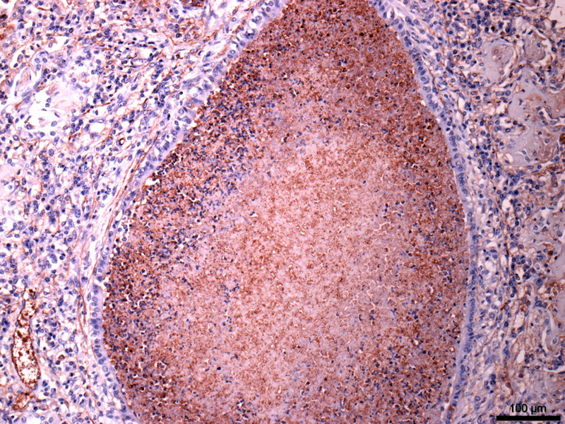
A bacterial pneumonia of multifactorial aetiology was confirmed in one of two six-week-old calves that had been vaccinated against PI3, BVD, RSV and Mannheimia at a calf rearing unit. Histopathology of the lung revealed severe, acute bronchopneumonia with intralesional bacteria typical of that seen with Pasteurella multocida, M haemolytica or Histophilus somni. Evidence also existed of subacute necrotising pneumonia, with extensive areas of necrosis bordered by degenerate neutrophils and mononuclear cells, suspicious of Mycoplasma bovis infection. This was confirmed on immunohistochemistry.

Several cases of lungworm in lambs have been identified on Baermann testing, primarily presenting with coughing. The most pathogenic species in sheep is D filaria. Protostrongylus rufescens may be pathogenic with heavy burdens and Muellerius capillaris is minimally pathogenic.
Maedi visna (MV) was detected in several flocks on serology. In one flock, sheep were purchased from a MV accredited flock and, after arrival on farm, inconclusive cases were reported on serology in the original flock. Subsequent screening of the purchased sheep detected one strongly seropositive animal. In another case, the main presenting signs were weakness, neurological deficits and weight loss, suggesting this was the visna form of MV.
Ovine pulmonary adenocarcinoma (OPA) was diagnosed on histopathology of lung samples from thin ewes on four occasions. In two cases, evidence existed of severe secondary bacterial pneumonia, a not uncommon finding in OPA cases.
Systemic pasteurellosis was the cause of sudden death in a six-month-old lamb. Necrotising pneumonia and hepatitis were seen on histopathology and Bibersteinia trehalosi was cultured from liver.
In a second case, two lambs died acutely and B trehalosi was isolated in mixed growth from congested lungs seen at postmortem. The lambs had been vaccinated twice against Pasteurella. In a further case, lung was received from 1 of 11 sheep in a flock of 200 to die over a two-week period. Histopathology revealed severe interstitial pneumonia compatible with systemic pasteurellosis; M haemolytica was isolated from lung.
Mycoplasma ovipneumoniae infection was confirmed on paired serology in a seven-month-old coughing, pyrexic Zwartbles lamb. M ovipneumoniae is often isolated from cases of ovine atypical pneumonia and may predispose animals to pasteurellosis and respiratory viral infections. Atypical pneumonia can be a particular problem in lambs housed in conditions with suboptimal ventilation. Histopathology of the lung is also a very useful means of determining the likely involvement of M ovipneumoniae infection in cases of pneumonia, with some cases showing almost pathognomic features for this agent.