9 Mar 2020
Systemic and miscellaneous diseases in winter 2019-20 are the focus on Axiom Veterinary Laboratories’ latest monthly update.
James Barnett
Job Title
Image © Uschi Dugulin / Pixabay
Presented are selected cases from the ruminant diagnostic caseload of Axiom Veterinary Laboratories.
Axiom provides a farm animal diagnostics service to more than 300 farm and mixed practices across the UK, and receives both clinical and pathological specimens as part of its caseload. The company is grateful to clients for the cases presented in this article.
The focus for this article is systemic and miscellaneous diseases in winter 2019-20.
Two calves that tested positive for bovine viral diarrhoea (BVD) on tag and testing as neonates were retested at four months of age by PCR.
One had a low cycle threshold (Ct) value (high level of virus) consistent with PI status, but the second had a much higher Ct value (very little virus) that was much more suggestive of transient infection.
BVD antigen ELISA run on the same sample from the second calf was negative, with a strong positive titre on serology; higher than one would expect for microbial detection array.
It was likely the PCR test was detecting viraemia left over from a transient infection, as reports exist in the literature of PCR picking up virus for more than 100 days post-infection.
BVD viraemia also was confirmed on PCR in a young calf – younger than three weeks old – with a domed head, hyperextension of the carpal and metacarpal joints, and hair loss; its twin was similar, but less severely affected.
Seven cases of malignant catarrhal fever were diagnosed this quarter – five on PCR and two on serology.
Clinical signs reported included:
A positive titre to maedi-visna was found in a five-year-old ewe with neurological signs, consistent with the nervous form of the disease, visna.
A previous case in the flock had been diagnosed on postmortem examination (PME) at the RVC.
A two-month-old Simmental calf died following a period of illthrift.
On PME, crusty yellowish white lesions were found on the tongue and white plaques on the hard palate. Histopathology revealed a hyperplastic and erosive glossitis with intralesional inclusions consistent with bovine papular stomatitis, although this was unlikely to explain the death of the calf.
Orf was confirmed by PCR in:
Orf also was confirmed on histopathology in an 18-month-old Texel ram with several granulomatous lesions on the top and bottom lips, with severe proliferative, necrotising dermatitis and intracytoplasmic inclusion bodies seen.
Salmonella enterica serovar Dublin was isolated from a faecal sample taken from a group of calves with pneumonia and, in one case, gangrene of the distal hindlimbs. The latter is a less commonly reported sequel to systemic Salmonella enterica serovar Dublin infection caused by endarteritis.
Salmonella enterica serovar Dublin was also isolated from joint fluid from a six-week-old suckler calf with joint ill.
Leptospirosis was suspected to be the cause of pyrexia and milk drop in two-year-old to three-year-old Holstein heifers, one animal seroconverting on paired serology.
Mycoplasma bovis was detected by PCR on a swab of joint fluid from an 18-month-old Limousin bull that had been bought in six weeks previously and had a history of multiple limb lameness, with visible swelling of the right hock, and the fetlocks of the left forelimb and left hindlimb.
A second case was confirmed in a herd with a short history of spontaneous joint swelling in dairy cows.
M bovis also was suspected to be the cause of severe, unresponsive lameness in four Holstein-Friesian dairy cows with swollen joints, after high positive titres were detected on serology in three of the animals and a moderate titre in the fourth.
Mycoplasma conjunctivae was detected by PCR in an outbreak of keratoconjunctivitis in housed ewes.
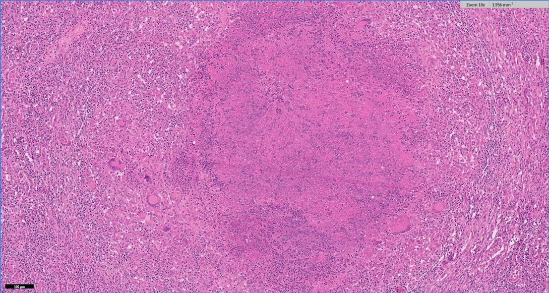
Mycobacterioisis was diagnosed on histopathology in a six-year-old sheep that was losing condition and, on PME, had multiple abscesses in the lungs, liver and mediastinal lymph node.
Granulomatous and necrotising pneumonia, hepatitis and lymphadenitis were present, and acid-fast bacilli were within foci of necrosis.
It was not possible to ascertain which mycobacterial species were involved; however, Mycobacterium bovis has been reported in sheep previously. Most cases are identified at slaughter, with only small numbers identified while still on-farm.
Mycobacterial culture on fresh tissue was recommended, and it was advised the local APHA office was contacted to discuss the findings and further testing.
Pulpy kidney was suspected to be the cause of six sudden deaths out of 400 non-vaccinated lowland lambs. Clostridium perfringens alpha and epsilon toxins were detected by ELISA in small intestinal contents.
Excess straw-coloured peritoneal and pericardial fluid, and small intestinal haemorrhages were found on PME.
Type D clostridial enterotoxaemia was suspected to be the cause of death in a Toggenberg goat, with the detection of C perfringens alpha and epsilon toxin by ELISA in faeces, where gross PME revealed an injected, septicaemic carcase and haemorrhage in the duodenal mucosa.
Streptococcus dysgalactiae was isolated from joint fluid from a one-week-old Suffolk-cross lamb with joint ill.
This is the most common cause of joint ill in young lambs and it is thought infection can arise during passage through the vagina, from oronasal contact with the ewe or from infected milk.
S dysgalactiae can survive on straw for extended periods, so entry via the navel is also possible. Other possible portals of entry are ear tagging and ringing.
Listerial encephalomyelitis due to Listeria monocytogenes was diagnosed on histopathology in a shearling, one of five sheep to die following signs including hyperaesthesia, collapse and falling to the left.
This is typically an ascending neural infection arising from the oral cavity and commonly associated with tooth eruption/loss.
L monocytogenes also was cultured from the brain stem of an eight-month-old lamb.
Six of eight suspected cases of caseous lymphadenitis (CLA) in one flock were positive on CLA serology.
In other suspected cases of CLA in three flocks, the isolation of Actinobacillus lignieresii from abscesses was consistent with a diagnosis of cutaneous actinobacillosis. Trauma usually allows the bacterium to gain entry. Streptococcus uberis also was isolated from a large abscess in the prefemoral lymph node of a milking goat.
Chorioptes bovis infestation was diagnosed in a north Devon suckler herd.
Sheep scab was diagnosed by microscopy in a number of flocks.
In one case, the only affected lamb in a group of 65 had severe pruritic, erythematous, crusting dermatitis. Staphylococcus chromogenes and Trueperella pyogenes were also isolated, and likely to be contributory factors.
In a second case, sheep scab was confirmed on skin microscopy in a five-year-old with crusted fleece coming off in clumps, despite two injections of ivermectin at a two-week interval.
Sheep from several flocks with signs potentially consistent with sheep scab had positive antibody titres, although a positive result does not confirm current infection and, in some cases, can be detected for up to nine months after successful treatment.
Fibropapilloma was diagnosed on histopathology in two bulls on different farms – one on the bridge of the nose, the other on the penis.
These tumour-like proliferations are associated with infection by a bovine papillomavirus and lesions typically resolve once adequate cell-mediated immunity develops.
Lymphoma was diagnosed on histopathology as the cause of a lump in the vaginal wall of a three-year-old Hereford heifer; this had a high mitotic rate, indicating malignancy, and the animal had a growth on the right mandible around the incisors.
Lymphoma was also the origin of nodules in the kidneys and enlarged lymph nodes in a Holstein-Friesian cow that had an right displaced abomasum and peritonitis on PME.
In this age of animal, the most common presentation is multicentric sporadic bovine leucosis, which is the result of spontaneous neoplastic transformation of lymphocytes. Lesions can occur at different sites, including the skin, lymph nodes, visceral tissues and, less frequently, periorbital sites. The condition can wax and wane, but is invariably progressive.
The possibility of enzootic bovine leucosis caused by enzootic bovine leukosis virus was raised as a differential, and it was advised the cases be reported to a local APHA office as they may have wished to undertake additional testing for this notifiable disease.
Mesothelioma was diagnosed on histopathology as the cause of multiple masses on the peritoneum, rumenal and abomasal walls in a five-week-old Belgium blue-cross calf; a large quantity of serosanguinous peritoneal fluid was also present.
These are neoplasms of body cavities and the peritoneum is the most frequent location in cattle; ascites is commonly seen and, if present before birth, can result in dystocia.
Bovine peritoneal mesotheliomas typically metastasise to regional lymph nodes.