21 Aug 2020
Cases of reproductive and respiratory disease from spring and early summer 2020 are the topic of Axiom Veterinary Laboratories’ latest update.

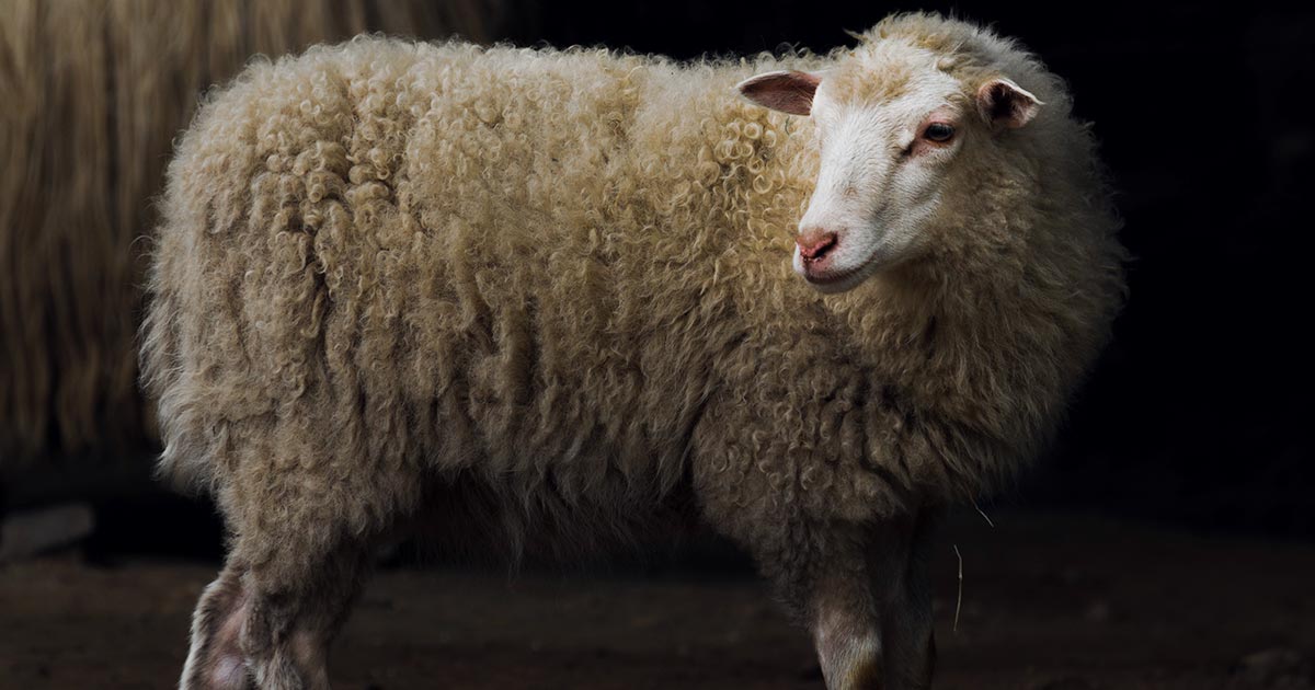
Image © Alexas_Fotos / Pixabay
Presented are selected cases from the ruminant diagnostic caseload of Axiom Veterinary Laboratories.
Axiom provides a farm animal diagnostics service to more than 300 farm and mixed practices across the UK, and receives both clinical and pathological specimens as part of its caseload. The company is grateful to clients for the cases presented in this article.
The focus for this article is respiratory and reproductive disease in spring and early summer 2020.
Several cases of infectious bovine rhinotracheitis (IBR) were confirmed on PCR. These included:
An IBR breakdown was suspected in a dairy herd after the bulk milk sample became antibody positive having previously been antibody negative. Follow-up testing on six cows confirmed this was the case as four were antibody positive for IBR virus.
Respiratory syncytial virus (RSV) was diagnosed, by PCR of the lung, as the cause of the pneumonia that had caused the relatively sudden death of a three-month-old Charolais calf at grass, in a herd in which two recent similar cases had occurred.
The calf had been treated with oxytetracycline, but died the following day. Histopathology found evidence of bronchointerstitial pneumonia consistent with a viral aetiology.
RSV was detected in three further cases on PCR testing:
Active seroconversion to RSV was seen in three of four unvaccinated four-month-old weaned Friesian calves in an acute outbreak of respiratory disease in one herd, and in 8 of 11 RSV-vaccinated Holstein-Friesian calves in a respiratory disease outbreak in a second herd. In the second case, one calf also seroconverted to bovine viral diarrhoea (BVD).
Parainfluenza-3 (PI3) was detected on a swab from one of four two-month-old calves with serous nasal discharge and pyrexia (temperature higher than 39.5°C).
A number of cases of ovine pulmonary adenocarcinoma, often with secondary bacterial infection, were diagnosed on histopathology, with the latter occasionally on bacteriology.
Seroconversion to maedi-visna was reported in a number of flocks, including one in which sheep were dying after a two-week period of noticed condition loss, with three of four animals tested being seropositive.
Several cases of bacterial pneumonia were reported in cattle. Some cases were diagnosed on histopathology, occasionally with culture; others were diagnosed on multiplex PCR.
Bacterial pathogens can act as primary pathogens; however, other predisposing causes – such as stress, management changes, adverse environmental conditions and concurrent immunosuppressive disease (such as BVD and tick-borne fever) are potential predisposing causes.
Organisms such as Pasteurella multocida, Mannheimia haemolytica, Histophilus somni and Trueperella pyogenes are likely to be implicated, and these were detected primarily using multiplex PCR as opposed to culture. Multiplex PCR does not rule out previous viral involvement in chronic cases, as any viral antigen originally present is likely to have been eliminated.
Culture did prove useful in identifying two cases of H somni pneumonia in a six-week-old Limousin-cross male calf with extensive pulmonary consolidation, and in two-week-old to five-week-old calves in a dairy herd; here, histopathology confirmed a bacterial aetiology to the pneumonia.
Cases of bacterial pneumonia were particularly common in young calves in both dairy and suckler herds, but occasionally reported in adult animals – for example, a housed dairy cow that died following peracute pneumonia where M haemolytica was isolated from the lung.
Pneumonia due to Mycoplasma bovis was confirmed in a two-month-old Aberdeen Angus calf with characteristic bronchointerstitial pneumonia, with caseous and coagulative necrosis on histopathology and M bovis detected by PCR on a lung swab. Although no clear evidence of an underlying viral cause existed on histopathology, PI3 also was detected by PCR.
M bovis also was detected with other bacterial pathogens on multiplex PCR – and very high positive titres to M bovis – in four-month-old unvaccinated Holstein-cross calves in a long-standing bovine respiratory disease outbreak.
An unusual case of pneumonia – with club colony formation in the lungs – was diagnosed in an eight-week-old suckled calf, which was a poor doer and had been pyrexic and unresponsive to treatment. It had an abomasal ulcer, as well as ulcers on the lips, buccal region and sublingually.
Testing for BVD antigen using ear tissue was negative. On histopathology, the findings were of a moderately severe diffuse subacute interstitial pneumonia and of focal granulomatous pneumonia with club colony formation, which is associated with certain species of bacteria such as Actinobacillus, Actinomycetes and Staphylococcus.
It was suspected to have been a haematogenous bacterial insult and the mouth lesions may have been the primary site of infection in this case.

Pneumonia due to M haemolytica – and, less frequently, P multocida – was diagnosed on culture in several flocks.
The affected lambs ranged from 1 week old to 12 weeks old, were reported as sudden deaths, acute pneumonia or fading lambs, and PME findings were of consolidated lung lobes with or without fibrinous pleurisy.
In cases where histopathology was carried out, acute suppurative and necrotising pneumonia was seen.
One flock had multiple lambs with joint ill and concerns over tick pyaemia. A 12-week-old lamb that had died in this flock was found to have fibrinous pleuropneumonia and M haemolytica was isolated from the lung despite having completed a vaccination course against pasteurellosis three weeks earlier. No significant organisms were isolated from the joint.
Chronic bronchointerstitial pneumonia with marked bronchiolar epithelial hyperplasia, consistent with atypical pneumonia, was diagnosed in a number of lamb flocks. This has a multifactorial aetiology – potentially including P multocida, M haemolytica, Bibersteinia trehalosi, Staphylococci, Streptococci and Mycoplasma ovipneumoniae.
The condition is seen most frequently in post-weaned lambs that present with ill thrift and a mild cough, although some will present as sudden death associated with a secondary fulminating pneumonia.
Infection can be more likely to occur if lambs are housed in early life in overcrowded, poorly ventilated sheds.
In one flock, histopathology typical of atypical pneumonia was found in a thin ewe, one of several to have died in the flock in recent weeks. Interestingly, evidence also existed of significant parasitic pneumonia with organism morphology most suggestive of either Muellerius capillaris and/or Protostrongylus rufescens. Both of these are common parasites of sheep and do not normally result in significant pneumonia; however, the combination of bacterial infection and a heavy parasite burden in this case was believed to be significant.
Salmonella enterica subspecies diarizonae serovar 61:-:1,5,7 – a sheep adapted strain of Salmonella known to cause chronic proliferative rhinitis in sheep – was isolated from a nasal swab taken from a ram with a six-week to eight-week history of bilateral mucopurulent nasal discharge.
Three weeks of antibiotics did not clear the infection, which can result in significant sized polyps in the nasal passages.
Usually, multiple cases exist in a flock – and it was subsequently isolated from a 12-year-old ewe in the same flock that was found collapsed, pyrexic and showing signs of respiratory distress.
Dictyocaulus viviparus lungworm infestation was diagnosed on Baermann testing and serology in a number of herds. Where a clinical history was given, coughing was unsurprisingly the most common presenting sign reported.
Muellerius capillaris lungworm infestations were detected in two goat herds with a history of coughing in adult goats. This parasite tends to be more pathogenic in goat herds than in sheep flocks.
Bacillus licheniformis was the cause of abortion in three suckler herds.
In one case, 6 out of 100 cows had aborted. It was also the cause of abortion in a heifer in a Holstein dairy herd where two abortions had occurred.
Infection is usually associated with the feeding of poor-quality silage and the organism has been shown to build up in water troughs.
Listeria monocytogenes was isolated from fetuses aborted in two Holstein herds and from a fetus in a Holstein-Friesian herd that had between 5% and 8% of stillbirths and abortions, and where one seven-month-stage fetus was noted to have fibrin on the pericardium.
It was also isolated from fetuses aborted in three flocks where multiple abortions were occurring.
Listeria ivanovii was the cause of abortion in an ewe in a flock where approximately 100 ewes had aborted and with a high barren rate at scanning.
Trueperella pyogenes was the cause of an abortion in a dairy herd. This tends to be a sporadic cause of abortion.
S enterica serovar Dublin was the cause of the abortion of a dairy cow at seven months to eight months of gestation, while Salmonella 6,7:-: enz15 was isolated from the aborted five-month-stage fetus from a Holstein-Friesian cow.
Enterococcus faecium was isolated in pure growth from an aborted dairy fetus. Although an enteric commensal, like Escherichia coli, it may have the ability to cause abortion through opportunistic invasion of the gravid uterus.
Vibrio furnissi was isolated in pure growth from an aborted fetus in a dairy herd.
This bacterium is typically associated with aquatic, primarily marine environments and is an emerging human pathogen.
Its role in the abortion was unclear and the cow was known to be seropositive for Neospora, but it was a zoonotic concern.
Several cases of enzootic abortion were diagnosed on the basis of modified Ziehl-Neelsen smears of placentae – in some cases backed up with Chlamydia abortus PCR – with abortions occurring within three weeks of lambing where reported.
In one flock, primarily stillbirths were reported and, in a second flock, both weak, fading lambs and abortions were present.
In one upland flock that vaccinated replacements against toxoplasmosis, concurrent Toxoplasma infection also was detected by PCR.
Toxoplasma-associated abortion was diagnosed through PCR testing of fetal placentae and brain, and fetal fluid serology in a number of flocks. In one ewe from a flock in which 5 of 370 ewes had aborted over a two week period, Bacillus cereus also was isolated from fetal stomach contents; this is a known abortifacient in cattle, but its role as an abortifacient in sheep is less well documented.
Seroconversion to Toxoplasma was seen in several flocks with abortion/barren percentages as high as 20%.
Three cases of abortion involved Salmonella diarizonae variants and two cases of of E coli and Campylobacter fetus occurred. In one of the C fetus cases, the abortion was in a two-year-old ewe – 1 of 20 abortions over a three-week period that occurred in two waves. The farm bought in mules yearly, and predominantly twins and triplets were affected. One case of abortion associated with L monocytogenes also was identified.
A heavy growth of an Aspergillus species was isolated from the fetal stomach contents of an aborted fetus, consistent with a diagnosis of mycotic abortion. Typically, this is caused by the ingestion of mouldy feedstuffs.
A Malbranchea species mould was isolated from a five-month stage aborted fetus in a dairy herd. It can be found in decaying vegetation, soil and animal faeces.
Neospora was suspected to be the cause of abortion in several herds – with positive results on maternal and fetal fluid serology, and PCR testing of fetal brain and placenta – although histopathology is required to confirm neosporosis as the cause of an abortion.
In one suspected case, a three-day-old south Devon calf with ataxia since birth was born into a herd with a history of Neospora exposure. The calf was negative for BVD antibodies and antigen, but had a high level of antibodies to Neospora. These could have been due to congenital infection or colostral antibodies, but even with the latter a very high risk would still exist of it being congenitally infected.
While many infected calves are born normal, in some cases clinical signs – such as ataxia, paralysis and hyperextension of the forelimbs or hindlimbs – can occur.
Neosporosis was also suspected to be implicated in a spate of recent abortions in a dairy herd, with six out of seven recently aborted cows being seropositive and a high positive titre being detected in a bulk milk sample.
Evidence of BVD virus infection was detected in abortion cases from a number of herds on either PCR or antigen ELISA testing.
In one case, where a five-year-old cow had aborted at five months of gestation, the history was of several cows having delivered weak calves or the calves having died.
Investigation of a six-month-stage abortion in a dairy herd – in which it was suspected active BVD virus infection existed and where salmonellosis had been diagnosed recently – a level of BVD virus in the foetus was detected that was suggestive of it being a persistently infected animal. BVD virus infection may, therefore, not have been the cause of the abortion, but it confirmed active BVD virus infection was present in the herd.
Evidence of Border disease viraemia or exposure was detected in several flocks where the histories included findings such as a high barren rate; stillbirths; small, weakly lambs being born; or lambs that were suspected to be “hairy shakers”.
In a 1,200-ewe flock, 50% of the yearlings had been barren, 10 of 10 ewes sampled had antibodies to border disease virus and a hairy shaker lamb was identified on PCR testing.
High positive titres to Schmallenberg virus were detected in two five-year-old to six-year-old suckler cows that had deformed calves at term, indicative of previous exposure to the virus.
Antibodies can remain detectable for years following infection – for a definitive diagnosis, calves need to be tested ideally for both the virus (for example, PCR on the umbilicus) and for antibody (on fetal fluid/serum).
The less common mastitis pathogens isolated on culture in cattle included Aerococcus viridans, Bacillus cereus, Candida species, Enterococcus saccharolyticus, Lactococcus garvieae, Lactococcus lactis, L monocytogenes, P multocida, Serratia liquefaciens, Serratia marcescens, Streptococcus canis and Trueperella pyogenes.
Lactococcus species are emerging mastitis pathogens and can cause clinical, subclinical and persistent high cell counts. They tend to be at a higher incidence in summer and autumn. Cure rates are often low with L lactis (approximately a third of cases) and repeat cases are not uncommon.
P multocida is usually a commensal in the oral cavity and upper respiratory tract; haematogenous spread also could be the route of transmission to the udder under certain conditions and suckling calves may be a source of infection.
M bovis was detected in cases of intractable mastitis refractory to treatment in a number of herds.
In one herd, a history existed of joint problems in cows and calves, respiratory disease in calves, and mastitis cases where little milk was expressed and cows became systemically ill.
M bovis was detected by PCR in a milk sample from one cow, a second cow with acute mastitis actively seroconverted to M bovis on paired serology, and a high titre – likely to be consistent with recent infection – was seen in a third cow with chronic bursitis over one hock.
The two most common isolates from cases of mastitis in sheep were M haemolytica and Staphylococcus aureus.