15 Jan 2018
James Dixon discusses diagnosis and management options for this condition, with a focus on the more common complaints.
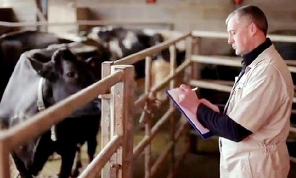
James Dixon
Job Title
The author on a farm with cattle.
As vets in practice we are often presented with individual lame cows, either on a first opinion basis, or because a particular cow has not responded to a previous treatment by the farmer or hoof trimmer.
These cases can often prove challenging, especially to recent graduates or someone unfamiliar with cattle foot trimming. This article will offer some suggestions on how to appropriately diagnose and treat some of the more common lameness presentations.
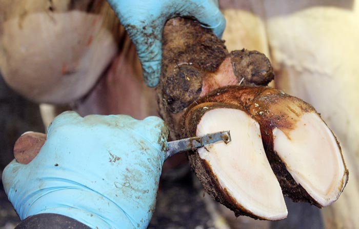
The correct equipment is critical when doing any job and hoof trimming is no exception. Panel 1 contains a basic toolkit the author would consider essential for anyone intending to pick up a cow’s foot.
To inspect the foot, it must be lifted and restrained safely – without causing undue discomfort to the cow. Most larger farms have good facilities for this, with a number using grant aid schemes to purchase electric or hydraulic foot crushes. Others may have more traditional “wopa box” style crushes, which can also work well.
Where possible, you should avoid makeshift systems, such as an old halter looped around a bar, as these are both dangerous and unlikely to give you the restraint required to do a decent job. If no facilities are available, it is worth knowing the telephone number of a good local hoof trimmer who may be able to bring a mobile crush to provide adequate restraint.
It is beyond the scope of this article to go through the full Dutch method of foot trimming, but, as a quick refresher, the steps are listed in Panel 2.
These steps provide the foundation of lame cow treatments and are used almost universally throughout the industry. The author encourages all farm animal vets to take an approved foot trimming course and show an interest in picking up the feet of lame cows, as it is an excellent way to show your enthusiasm to clients and engage with them on the subject of improving cattle mobility.
Since the advent of the Agriculture and Horticulture Development Board (AHDB) healthy feet programme several years ago, we have become familiar with the four success factors for controlling lameness and the most important, in the terms of this article, is: “early lameness detection and prompt, effective treatment”.
This simple statement is, in the author’s opinion, the single most important factor in determining the prevalence of lameness on an individual farm and should be the first thing we strive to improve when advising on cattle mobility. A great body of work exists showing how important early detection is, with cows treated quickly showing significantly improved cure rates versus those lame for longer. This means client education is key to try to break the cycle of continually focusing on chronically lame cows not likely to cure, when efforts may be better concentrated on those with a greater chance of success.
Herd mobility scoring is still the most reliable method of lameness detection and, if done frequently enough (every two weeks is recommended), it can be an excellent tool for the detection of new lameness cases and allow them to be treated as soon as possible.
Some cow activity monitoring systems are also starting to include lameness detection in their software algorithms and, while these are not all tried and tested yet, they will at least provide a flag that draws attention to a particular cow, so she can be assessed for lameness or other health issues.
The next component is effective treatment and this can only be possible with an accurate diagnosis, so the ability to correctly identify lesions is critical. Again, the AHDB healthy feet project has an excellent resource in its “foot lesion picture card”, which can be downloaded from the website (http://bit.ly/2Buz3zY).
Lameness lesions are split into three main categories:
Step 1 – trim the example claw (usually the inner claw on a hind foot and outer on a front foot) to the correct length (75mm to 85mm depending on the cow) then, sparing the heel, trim the sole to leave a 5mm step at the end.
Step 2 – repeat Step 1 with the treatment claw and trim to match the example.
Step 3 – model out a small area of sole toward the back of the example claw, and a larger area for the treatment claw to alleviate the sole ulcer site.
Step 4 – if any lesions have been identified, remove them from weight-bearing by lowering the sole, if possible (caudal 2/3 only), and applying a block.
Step 5 – Removal of loose horn, particularly at the heel where heel horn erosion can occur, and any under-run horn near to a lesion.
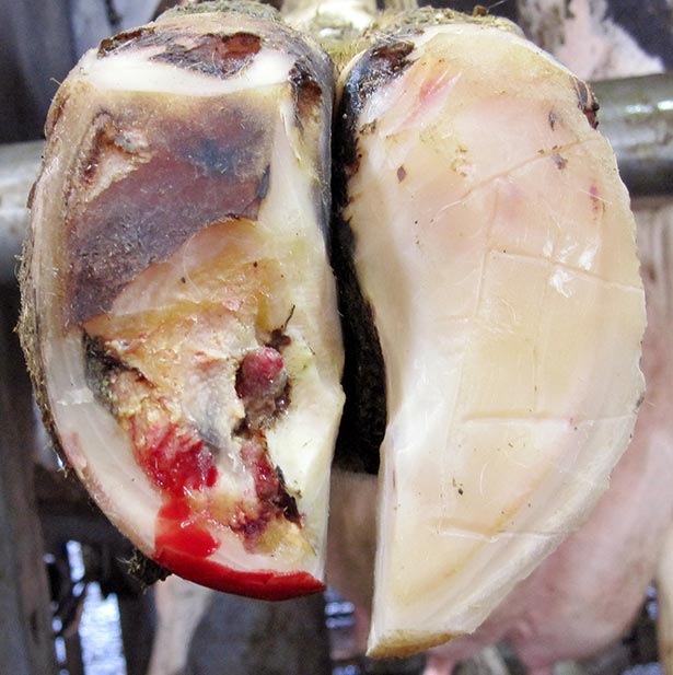
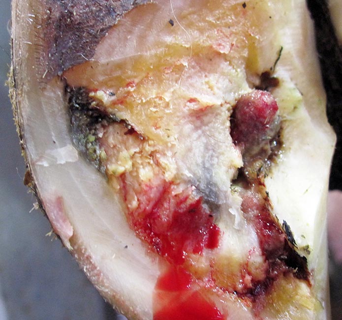
Sole haemorrhage and sole ulcers are essentially a progression of the same condition, as the haemorrhage is seen most often at the sole ulcer site. In most cases, the condition has progressed to the ulcer stage before the cow becomes overtly lame, so this is what is seen when trimming.
Research has shown the best cure rates for these conditions are achieved when a five-step trim is combined with a block and NSAID. This should be the gold standard treatment for any lame cow presenting with sole haemorrhage or an ulcer, and is particularly important early on in the disease process, as it is only by applying the correct treatment here we can prevent a more chronic lameness, which will lead to complications such as secondary infections and bone changes on the surface of the pedal bone that will predispose to further cases of lameness in the future.
Antibiotics are not normally indicated in cases of sole ulcer, but, if a more chronic lesion is present, a risk of secondary infection exists either to the tendon sheath or the distal-interphalangeal joint. In cases of a chronic ulcer with swelling and erythema in the heel area, a course of antibiotics would be indicated, with five to seven days of penicillin the preferred choice along with NSAID therapy.
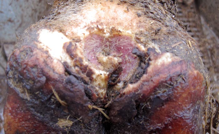
The key principles of treatment for WLD are the same as sole ulcers, with a thorough five-step trim, a block and treatment with NSAIDs. However, due to the fact an infection is almost always present in a case of WLD, a need exists for a particular focus on step four of the Dutch method – removal of loose horn.
This should be done carefully and with a very sharp hoof knife, with the objective of removing any horn not firmly attached to the hoof without cutting into healthy tissue and drawing blood. This can be difficult, especially in acute cases with active inflammation and the area around the infection still very vascular. It is often prudent to relieve the initial pressure, apply a block then suggest a re-examination in two to three days, when more of the loose horn can be removed to ensure a rapid recovery.
WLD lesions can track up the hoof wall as well as under the sole and it is important to follow any track up here as well, sometimes removing a whole section of wall. These will heal very effectively so long as the affected claw is removed from weight bearing with a block and all the loose horn has been removed.
DD is a bacterial condition caused by a group of spirochaetes called treponemes. It can present in either an acute or a chronic form and is often graded using the “M” system, which has been described before by Pedersen (2017).
In terms of treatment, many different products are available, but the key to success is in effective cleaning and debridement of the lesion, followed by repeated application of a suitable topical product. The author recommends cleaning with dry cotton wool, which is abrasive and will remove a lot of the hyperkeratotic parts of the lesion that can prevent the topical treatment from getting to the site of infection, which can be quite deep.
Once cleaned and debrided, good results have been demonstrated using topical tetracycline sprays, or a non-antibiotic alternative containing chelated copper and zinc, which the author thinks are as effective. It is recommended treatment is repeated daily for five to seven days to maximise the chances of achieving a cure.
Interdigital necrobacillosis is generally seen as an acute condition, often causing severe lameness. The foot is swollen above the coronary band and has a moist area in the inter-digital space with a characteristic “foul” smell.
Treatment is with a routine foot trim to rule out other conditions, cleaning and spraying the affected area then a short course of penicillin, usually accompanied by an NSAID to reduce the swelling and associated pain.
If not picked up quickly enough, or if combined with a DD infection, these lesions can be particularly aggressive and quickly track into the foot and involve the proximal interphalangeal joint. In these cases, a digit amputation may be the only treatment option available.
Non-healing lesions have become more common in recent years as the prevalence of DD rises within the national herd and studies have consistently demonstrated the presence of the DD treponemes in these lesions.
They include conditions such as toe necrosis and wall ulcers, and, while the term “non-healing” can be discouraging, they can be treated successfully, provided they are picked up early and the treatment is both aggressive and sustained to ensure resolution.
The key principles of treatment for these lesions are as follows:
The key to success with these lesions is to be very thorough with the initial trim, but also to re-examine them on one to two further occasions to ensure they are progressing correctly. This should be discussed with farmers beforehand to ensure they are willing to invest sufficiently in the animal, and the treatment should be targeted at younger animals likely to remain in the herd for some time.
As stated previously, early lameness detection and prompt, effective treatment are key to success in treating lame cows, and the take-home message from this article should be to encourage both vets and farmers to work hard on this area through mobility scoring. Also, invest in suitable facilities to allow lame cows to be examined quickly and safely as soon as they are detected.
The Dutch five-step method provides an excellent framework for assessing and trimming the foot, but the secret to success also lies in proactive treatment with blocks and NSAIDs for the claw horn lesions, and aggressive cleaning/debridement and sustained therapy for infectious lesions, such as DD.
As the focus on lameness and cow welfare continues to grow, the author hopes the profession can take an active role in the control and prevention of lameness, but also in the rapid and successful treatment of individual cows.