24 Jul 2017
A look at the types and preventive steps regarding common diseases that affect normal metabolism of cows.
Sophie Mahendran
Job Title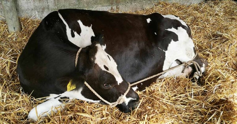
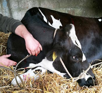
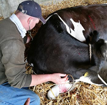
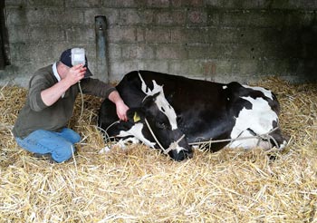
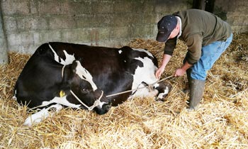
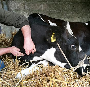
A cow restrained ready for calcium administration.
Most metabolic diseases of the modern bovine – with the exception of grass staggers – occur within the first two weeks postpartum, with the risk period extending to the time of peak lactation due to the high turnover and variable intakes/excretion of feed and mineral components in both beef and dairy cattle.

The economic consequences of metabolic disturbances, and long-term consequences of reduced performance and increased risk of culling, mean prevention of these management-related diseases is paramount for successful farming businesses.
The common metabolic diseases experienced by cattle are hypocalcaemia, ketosis, hypophosphataemia and hypomagnesaemia. However, the precursor to many of these can be traced back to the transition cow period (with the exception of hypomagnesaemia). Work has demonstrated distinguishable behaviour patterns in transition cows strongly related to an increased risk of metabolic disease post-calving, with these behaviours including decreased time at the feed bunk, along with fewer visits to it, and decreased movement (Belaid et al, 2016).
This demonstrates the long-term implications of management practices in the transition period, and highlights how far back vets may need to look to help identify the cause of metabolic problems when they arise in the lactating animal.
Hypocalcaemia – more commonly referred to as milk fever – primarily occurs around the time of colostrogenesis and initiation of lactation, with up to 50% of third lactation cows experiencing subclinical hypocalcaemia (Reinhardt et al, 2011). This same work also highlighted the surprisingly high prevalence of 25% subclinical hypocalcaemia in first lactation heifers.
Research over the past decade has led to milk fever being recognised as a gateway disease due to its immunosuppressive effects. Calcium acts as a second messenger through its release into the cytosol from intracellular stores in response to stimulus.
This typically reduces muscle contractility, leading to teat sphincter weakness and, therefore, mastitis; reduced abomasal contraction and, therefore, displaced abomasums; and slowed uterine involution, thereby causing slowed return to cyclicity.
Hypocalcaemia has also been demonstrated to increase cattle cortisol levels, thereby contributing to immunosuppression, resulting in decreased pathogen recognition by blood mononuclear cells and decreased neutrophil activity. Cows’ blood contains approximately 3.5g of calcium, but during the last trimester of pregnancy, the growing fetus requires approximately 12g/day, followed by an increase to 20g/day to 30g/day at the onsetof lactation (Goff, 2014).
This huge increase in calcium requirement is met by reducing calcium excretion in the urine, increasing calcium release from the bone fluid and matrix via osteoclast activity, and absorption of calcium from the diet by passive and vitamin D-dependent transport mechanisms. These processes can be inhibited by high potassium and low magnesium levels.
Clinical signs of hypocalcaemia can be split into three stages (Panel 1). Initially, tetany occurs due to low extracellular calcium, causing increased permeability of neuronal membranes to sodium ions. This causes a progressive depolarisation and less excitation required to open voltage gated sodium channels, which increases the possibility of action potentials.
Stage one
Stage two
Stage three
If the plasma calcium ions decrease to less than 50% of the normal value, action potentials may be spontaneously generated, causing contraction of peripheral skeletal muscles. However, this early stage of the condition is not frequently seen or recognised, with a more typical clinical picture being of a recumbent animal exhibiting flaccid paralysis.
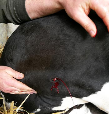
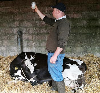
Treatments given at the occurrence of clinical signs of hypocalcaemia include the administration of IV and SC calcium, with additional oral calcium in the form of a drench or bolus (although this only boosts blood calcium levels for 6 to 10 hours). IV administration should be conducted after the solution has been warmed to body temperature, with auscultation of the heart to assess for arrhythmias, and is generally followed by administration of a further bottle SC.
Methods to prevent milk fever begin in the transition period, with the use of dietary anion-cation balance (DCAB) diets being fed for two to three weeks prior to calving. These diets contain low potassium and sodium, whose alkalinising effects are thought to alter parathyroid hormone (PTH) receptor structure. DCAB diets also have additional chloride and sulphate anions added to it to decrease blood pH, which restores tissue responsiveness to PTH. The net effect of the DCAB diet is slow reabsorption of calcium by the kidneys and promotion of upregulation of osteoclasts to begin the process of calcium mobilisation from bone reserves.
Ketosis is a condition that arises due to a mismatch in the energy requirements and intakes, leading to an overall negative energy balance. Approximately 30% of body condition scores are lost in the first 10 days post-calving, with fresh cows using their own body protein to support the amino acid and glucose requirements of milk production (Roche et al, 2007). Risk factors for ketosis include having twins, a high body condition score and a high milk production in the previous lactation.
High body condition scores (greater than 3.5) result in more non-esterified fatty acids (NEFAs) being released from adipose tissue, along with a greater decline in dry matter intake post-calving. Cows with subclinical hypocalcaemia also mobilise more body fat. Accumulation of the released triglycerides within the liver can cause dysfunction (fatty liver) and reduces the organ’s capacity for gluconeogenesis, increasing the ketosis risk. This process can be exacerbated by cows consuming excess energy pre-calving asthis prevents the release of NEFAs from adipose stores, which normally act to prime the tissues, such as the liver, for NEFA oxidation post-calving.
Negative energy balance and ketosis are also associated with prolonged immunosuppressive effects, resulting in decreased acute phase protein production and
reduced expression of cofactors needed for effective lymphocyte function (α-interferon and interferon-λ). The low protein levels experienced by many cows also result in low immunoglobulin production, with low levels of the amino acid glutamine being further linked to reduced immune function due to its role as an energy sourcefor white blood cells.
Treatment of ketotic cows begins with their identification through use of either urine or blood tests for identifying β-hydroxybutyrate (BHB). These can be
routinely used during fresh cow checks as not only an individual animal test, but also as a herd monitoring tool to assess the success of transition management. Cows identified as ketotic (BHB greater than 1.2mmol/L to 1.4mmol/L; Shin et al, 2015) can be treated with propylene glycol drenches, with the addition of a corticosteroid injection, if appropriate.
In addition, supportive management – such as provision of easy-access, palatable food and a low-stress environment – can be vital to ensure both recovery and continued maintenance of a positive energy balance.
Prevention strategies for ketosis begin during the previous lactation, with cattle being dried off at an appropriate body condition of 2.5 to 3, and this being maintained during the dry period. Further management of “at-risk” animals, such as those carrying twins, can then include use of monensin boluses and earlier movement into transition groups.
Maintenance of dry matter intakes should be encouraged towards the end of gestation, with low stocking densities and easy-access feeds that prepare the rumen for effective digestion of the milker’s ration.
Clinical hypomagnesaemia, commonly referred to as grass staggers, is associated with serum magnesium levels lower than 1.2mg/dl. Magnesium is important for normal nerve conduction and muscle function, along with being a cofactor for enzymatic reactions in every major metabolic pathway. Normal magnesium excretion is modulated through reabsorption from the kidneys, with little ability to mobilise bone stores in short-term deficits. As such, the diet is the major source of magnesium for cattle.
Cow diets should also contain suitable levels of bioavailable magnesium (dairy cows need approximately 0.4%), such as in the form of magnesium sulphate or magnesium chloride, which is soluble and, therefore, able to be absorbed across the rumen wall.
Active transfer of magnesium across the rumen wall requires use of a sodium-linked, adenosine triphosphatase-dependent transporter, and this process can be blocked by high potassium and nitrate levels, although passive transfer will still occur. Low magnesium levels also reduce PTH activity, which results in decreased calcium levels in the cow, which may explain many apparent mid-lactation milk fever occurrences.
Fresh spring grass is a major risk factor for hypomagnesaemia due to its low magnesium and sodium content, and high potassium levels. High potassium fertilisers also decrease magnesium uptake by plants. Clinical signs of hypomagnesaemia include twitching, hyperaesthesia, staggers, convulsions, increased heart sounds, increased heart rate and respiratory rate, and increased temperature. Treatment of hypomagnesaemia can be challenging if cattle are experiencing severe convulsions, so safety of both the vet and farmer should be considered. Great care must be taken when giving magnesium IV due to the possibility of inducing fatal cardiac dysrhythmias, medullary depression, respiratory failure and death.
Ideally, use a mixture of calcium and magnesium given IV, with additional magnesium given SC. Preventive intraruminal boluses can be given at risk periods to provide continuous release of magnesium, with magnesium oxide added to water troughsor applied as drench at a rate of 60g/day.
Phosphorus is maintained under homeostatic regulation through absorption from the diet and reabsorption from bone. It is used for bone and muscle growth, with late gestation fetuses requiring as much as 10g per day for growth. Blood phosphorus levels should remain above 1mg/dl.
Hypophosphataemia is strongly linked with hypocalcaemia, as the increase in PTH triggered by low calcium levels results in increased urinary and salivary loss of phosphorus. Along with this, high cortisol levels, such as those produced by high-stress events, also forces phosphorus into cells, thereby reducing its availability in the body.
It is quite uncommon to see cattle that are purely hypophosphataemic and it is generally always accompanied by dysregulation of calcium metabolism.
The main clinical presentation associated with hypophosphataemia is generally that of a recumbent cow that is otherwise bright and may still be eating, but unable to rise. Treatment of this condition generally focuses around correction of calcium levels, with additional use of oral phosphorus drenches.
Other phosphorus-containing products are available, but it is important to check they contain biologically metabolisable sourcesof phosphorus (PO4) for them to be used by the cow, and not PO2 or PO3, butafosfanor toldimfos, as these forms have to be converted to PO4 before they can be used (GrÜnberg, 2014).
Although the descriptions of these metabolic disorders has focused heavily around their occurrence in dairy cattle, all these conditions can, and do, affect beef cattle, too. The risk factors remain the same, although animals may not be detected until clinical signs are more advanced due to the lower level of human supervision these animals receive.
Preceding treatment of these animals, blood collection for biochemical analysis can be useful to confirm a diagnosis post-treatment or establish a diagnosis when initial treatment strategies appear to have been unsuccessful. This is especially true for the apparent down cow unable to rise following treatment for presumed metabolic disturbances.
Prompt treatment of these metabolic conditions often has a high clinical success rate, but the “silent” losses in the form of reduced productivity are often unmeasured and unappreciated. Farms suffering higher-than-expected levels of metabolic disease should be encouraged to invest in preventive strategies, with farm vets and nutritionists being key components of a successful farm team.