28 Nov 2023
Treating Dolly’s lesions: a surprising goat discovery
Author Matthew Smith details the case of Dolly the goat, whose lesions – thought initially to be a pyoderma – turned out to be something more serious…
Matthew Smith
Job Title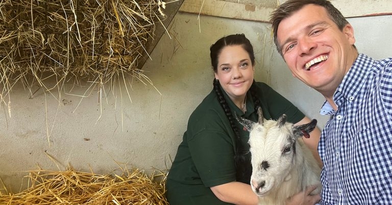
Dolly the goat with Matthew Smith, who treated her squamous cell carcinoma.
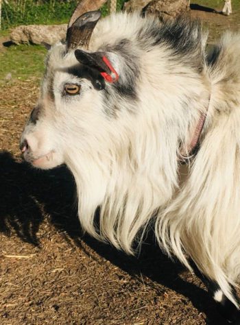
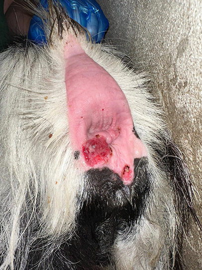
Dolly the goat (Figure 1) first presented to the author in December 2022 with scab-like lesions around her anus and perineal area that were red, ulcerated and had some purulent material coming from them.
Her condition was initially treated as though it was a pyoderma, with a course of amoxiclav antibiotic and anti-inflammatory meloxicam. After a week, limited resolution was present, so the course was continued with the addition of chlorhexidine wipes. This showed a mild improvement, but still no resolution.
Dolly was not itching the area, no other similar lesions were present elsewhere on her body and she had no issues with defecation. The next step was to take a swab for bacteriology culture and sensitivity. This did not yield anything other than normal skin flora and fauna.
The lesions seemed to worsen and become thicker over the next few weeks, with the only improvement from application of betamethasone cream.
Biopsy
It was decided that a biopsy was the next step and the results that came through were very surprising – a squamous cell carcinoma. The author was quite disheartened at this diagnosis, as he knew surgical resection was not possible.
The author read a report (Gibbons et al, 2015) that included a link to squamous cell carcinoma treatment with cryotherapy, to which he was elated, as all the equipment was on hand to attempt the treatment.
He researched the information and, using bottled gas nitrous oxide connected to a cryotherapy gun, the first session was planned.
Using a combination of xylazine and ketamine as anaesthetic and epidural local block, Dolly had four sessions of cryotherapy.
Method used
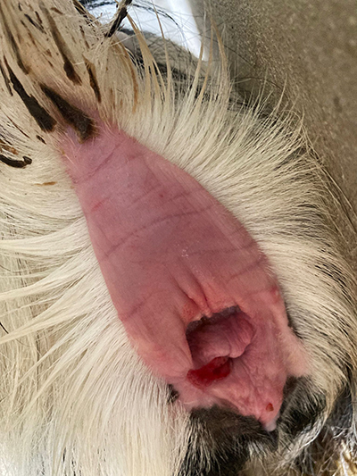
The author applied the gun with the disc attachment to the lesion for a quick freeze, 30 seconds on and then a slow thaw. He did this three times on the same area and moved to repeat it on a different part of the lesion as the carcinoma was so extensive (Figures 2a to 2e).
The paper said three cycles was enough to get rid of the cancers in their cases; however, given Dolly’s was so large and deep, he suspected it may take more than this.
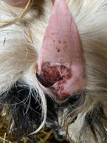
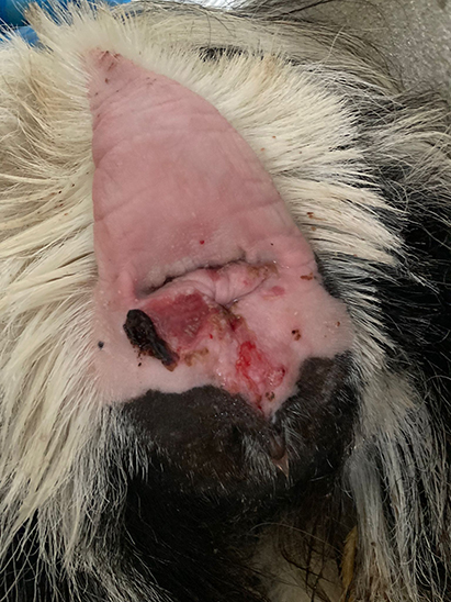
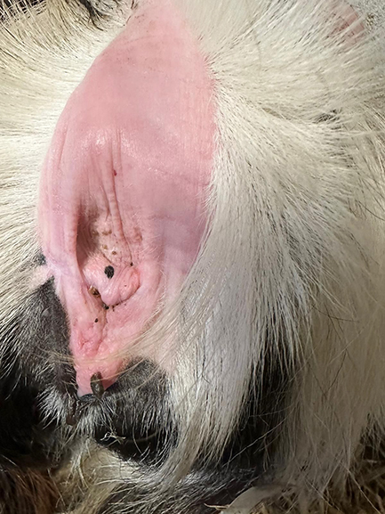
Her treatment schedule consisted of four parts on 16 March, 5 April, 2 June and 14 July. A final check was made on 12 September, showing the lesion had fully resolved.
- Use of some of the drugs in this article is under the veterinary medicine cascade.