Register

Login

Vet Times logo
+
  • View all news
  • Vets news
  • Vet Nursing news
  • Business news
  • + More
    • Videos
    • Podcasts
  • View all clinical
  • Small animal
  • Livestock
  • Equine
  • Exotics
  • All Jobs
  • Your ideal job
  • Post a job
  • Career Advice
  • Students
About
Contact Us
For Advertisers
NewsClinicalJobs
Vet Times logo

Vets

All Vets newsSmall animalLivestockEquineExoticWork and well-beingOpinion

Vet Nursing

All Vet Nursing newsSmall animalLivestockEquineExoticWork and well-beingOpinion

Business

All Business newsHuman resourcesBig 6SustainabilityFinanceDigitalPractice profilesPractice developments

+ More

VideosPodcastsDigital Edition

The latest veterinary news, delivered straight to your inbox.

Choose which topics you want to hear about and how often.

Vet Times logo 2

About

The team

Advertise with us

Recruitment

Contact us

Vet Times logo 2

Vets

All Vets news

Small animal

Livestock

Equine

Exotic

Work and well-being

Opinion

Vet Nursing

All Vet Nursing news

Small animal

Livestock

Equine

Exotic

Work and well-being

Opinion

Business

All Business news

Human resources

Big 6

Sustainability

Finance

Digital

Practice profiles

Practice developments

Clinical

All Clinical content

Small animal

Livestock

Equine

Exotics

Jobs

All Jobs content

All Jobs

Your ideal job

Post a job

Career Advice

Students

More

All More content

Videos

Podcasts

Digital Edition


Terms and conditions

Complaints policy

Cookie policy

Privacy policy

fb-iconinsta-iconlinkedin-icontwitter-iconyoutube-icon

© Veterinary Business Development Ltd 2025

IPSO_regulated

28 Jun 2022

Cytology findings from multiple skin nodules seen in a young dog

Francesco Cian presents the case of an 11-month Labrador retriever with wart-like aplopecic lesions in his latest Diagnostic Dilemmas column.

author_img

Francesco Cian

Job Title


Cytology findings from multiple skin nodules seen in a young dog

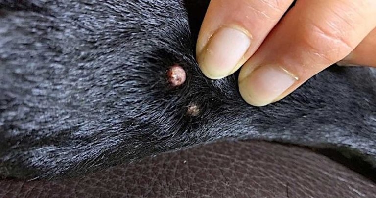
Figure 1. Nodules on the dorsal paw of the left hindlimb of a young Labrador retriever. Image: Emscote Vets

An 11-month-old Labrador retriever presented for recent onset of two small wart-like, alopecic lesions on the dorsal paw of left hindlimb (Figure 1). The veterinarian performed a fine-needle aspirate (FNA) of the lesions and submitted the slides to an external laboratory for cytological examination.

Figure 2a. Well-differentiated squamous epithelial cells (Wright-Giemsa 40×).

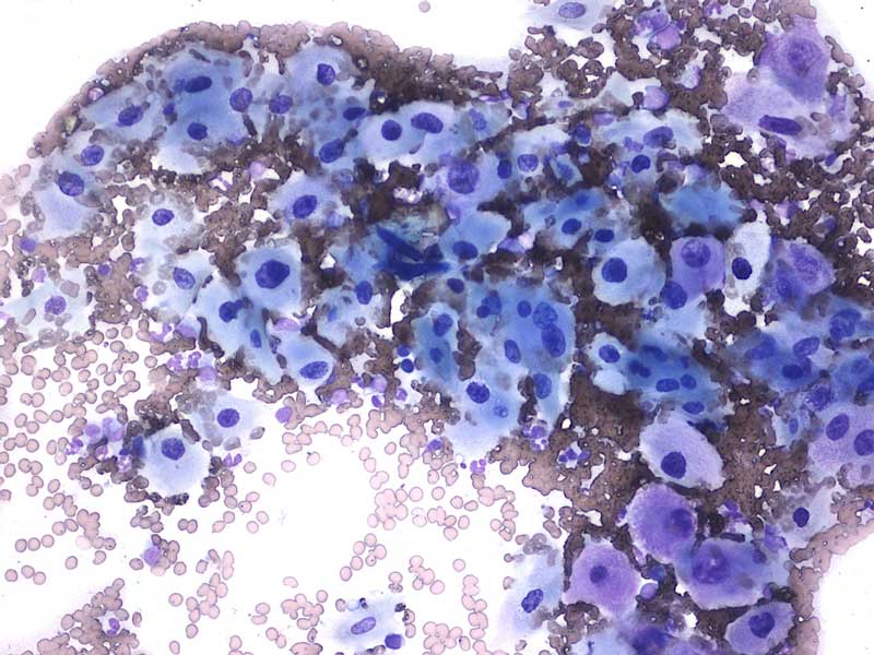
The aspirates from the two lesions (Figure 2) were similar and showed a main population of large squamous epithelial cells, either individualised or arranged in small groups, on a clear background with frequent red blood cells and few extracellular bacteria.

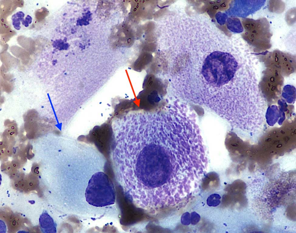
Cells appeared polygonal and angular, with low nucleus:cytoplasm ratio. Cytoplasm was abundant, pale basophilic (blue arrow) and contained bright fuchsia granulations in a small percentage of cells (red arrow). Nuclei were round, large and central to paracentral, with granular chromatin and poorly visible nucleoli. Few leukocytes, mostly blood-derived neutrophils, were also noted.

Diagnosis

The presence of a main population of large, well-differentiated squamous epithelial cells from a warty lesion of a young dog was consistent with papilloma.

Insight

Papillomas are classified as non-viral (squamous papilloma) and viral forms. The latter is caused by canine papillomavirus infections and has been associated with multiple syndromes: oral papillomatosis, cutaneous papilloma, cutaneous viral pigmented plaque, footpad papilloma and venereal papilloma.

Figure 2b. Well-differentiated squamous epithelial cells (Wright-Giemsa 100×).

Cutaneous papilloma is commonly observed in young dogs as single or multiple lesions. It presents as exophytic form, in which the folded epidermis protrudes above the surface of the skin, or inverted papilloma, in which the folded epithelium is contained within a depressed cup-shaped structure. Face, ears and extremities are preferred locations. Spontaneous regression within six weeks to nine months has been reported.