30 Jan 2017
Jerry Dunne describes a case in which a stray dog presented with a fractured spine after being involved in a road traffic collision.
Jerry Dunne
Job Title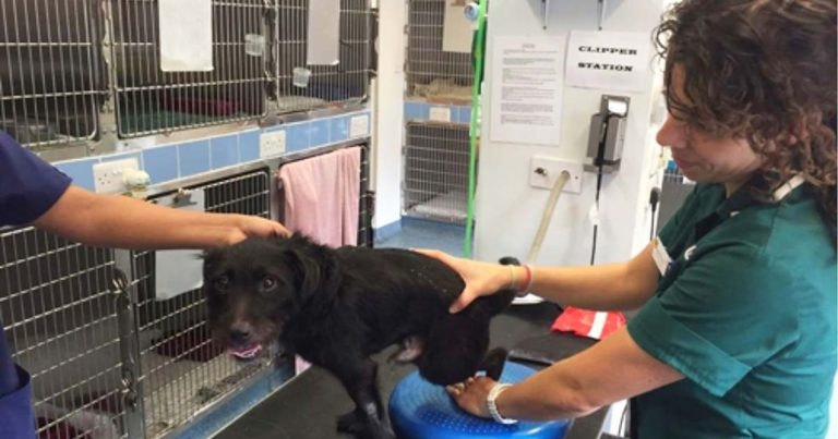
Paddy, a 14-month-old male Patterdale terrier cross stray, was hit by a car in South Ockendon, Essex. His owner was nowhere to be seen and the driver of the car did not stop.

The dog was left paralysed and in severe discomfort on the side of the road, but was spotted by a local resident and taken to Medivet South Ockendon for immediate veterinary attention.
Here, head vet George Vug and head veterinary nurse Karen Whittaker completed an x-ray, which revealed Paddy had a spinal fracture of the eleventh thoracic vertebral body. Because the veterinary practice was not open later than 7pm, the team transferred Paddy to Medivet Hendon 24 Hour Hospital to receive emergency treatment and around-the-clock nursing care.
Meanwhile, a photo of Paddy and a plea to find his owner was released on social media, with an overwhelming amount of support from the public flooding in and some offering to donate funds towards the cost of treatment.
While genuinely moved by the enormous public support, Medivet chief executive Arnold Levy said: “We really wanted to care for Paddy and, after seeing all the love pouring in for this pup, knew we had to give

him a chance and we were, without a doubt, going to foot the bill.”
Paddy arrived at Medivet Hendon with a stage IV spinal injury – deep pain was present, along with no proprioception, no voluntary movements, patellar reflexes and withdrawal reflexes present.
A CT scan was completed, which showed comminuted fracture of the eleventh thoracic vertebra, with at least eight fracture fragments, severe distortion of T11 and malalignment of the spinal canal, leading to severe compression and angulation of the spinal cord.
It was elected to perform surgery on Paddy, which involved right dorsolateral exposure of thoracic spine T9 to T13.
Multiple gelpies were applied to the dorsal spinal processes, facets and ribs of T10 and T12 to distract the spine and relieve compression forces on T11.

Once the fracture fragments were distracted to normal vertebral body length of T11, a hemilaminectomy was performed on T11. A combination of fragment removal, rongeurs and high-speed pneumatic drill were used to create a window to assess the spinal cord.
Blood clots were removed, as well as bone fragments within the canal causing compression to the cord.
A titanium locking plate was pre-contoured to be placed on the dorsolateral aspect of the thoracic vertebrae to buttress the fractured T11. Holes were drilled in the bodies of vertebrae T9, T10, T12 and T13, and locking titanium screws were placed.
The gelpies were removed and the spine checked for stability – good stabilisation had been achieved.
A postoperative CT scan was performed to check the screw placement and spine alignment. This revealed good purchase of screws in the vertebral bodies and decompression of the spine at T11.

Six weeks after the initial surgery, a second surgery had to be performed, as Paddy’s mobility began to deteriorate. He became ataxic in his hindlimbs with proprioceptive deficits. CT was repeated and two cranial screws were found to have become dislodged from T9 and T10 (Figure 1), with moderate callus formation at T11.
The plate was removed and contoured further for a better approximation to the spine (Figure 2), the original screws on T12 and T13 were replaced, new holes were drilled on T9 and T10 and new titanium screws were placed in these (Figure 3).
Postoperative CT showed good placement and alignment. A back brace was used for additional support as Paddy‘s activity levels had increased significantly since his original surgery.
An extended physiotherapy plan was developed for Paddy by veterinary nurse in MRI and CT Silvia Boscolo Contadin.

This physiotherapy plan involved basic massage, heat and shockwave therapy, as well as rehabilitation and physiotherapy to build up muscle on Paddy’s legs and improve his gait (Figure 4).
Eight weeks after the collision, a CT scan was performed, which revealed Paddy’s fracture had completely healed. He was regaining his strength and walking well with the support of his back brace.
Three months later, Paddy moved into his new home with Medivet student veterinary nurse Jodie Lester, who had been by Paddy’s side since he first visited Medivet South Ockendon (Figure 5).