8 May 2017
A Pancarpal arthrodesis Labrador retriever case
Simone Anesi and Toby Gemmill on the case of Shadow, a one-year-old, male neutered Labrador retriever that presented as an emergency after falling down a steep bank.
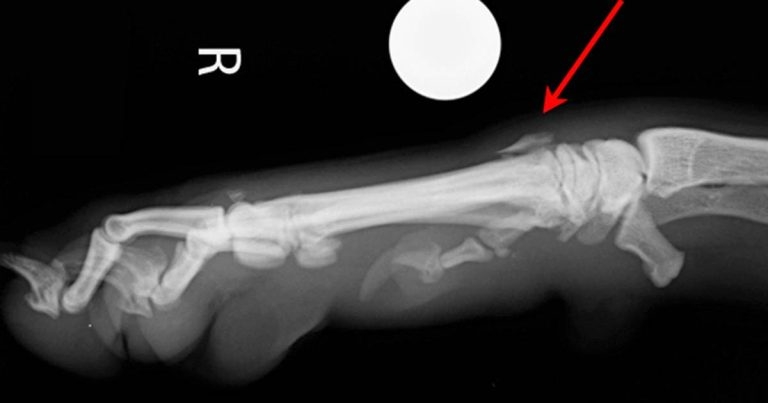
Figure 2. Mediolateral projection of the carpus showing metacarpal fracture (arrow).
Shadow, a one-year-old, male neutered Labrador retriever is presented as an emergency after falling down a steep bank, approximately 5m high.
On clinical examination he is very bright and reactive, although non-weight bearing lame on his right forelimb. The right carpus is swollen and painful; distal limb sensation and blood supply appear to be intact. The remainder of your clinical examination is unremarkable.
The patient is sedated and orthogonal neutral views of the carpus are obtained.
Question
What do the radiographs show?
Answer
On the dorsopalmar view (Figure 1) a fracture of the proximolateral aspect of the fifth metacarpal bone is present. There is also suspicion of a fracture of the first carpal bone (although further oblique views or advanced imaging, such as a CT scan, would be necessary to confirm the latter).
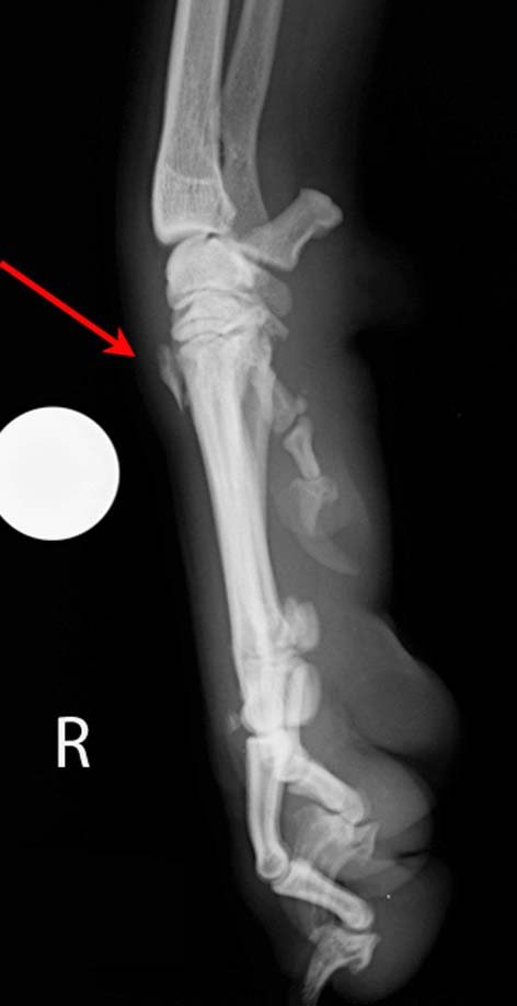
On the mediolateral view (Figure 2), a fracture of the proximal aspect of either the third or fourth metacarpal can be appreciated, but it is difficult to localise this lesion accurately as the fragment cannot be visualised on the dorsopalmar projection.
On examination of the patient under sedation, you can appreciate the right carpus has multidirectional instability. You proceed to obtain mediolateral and dorsopalmar stressed radiographs.
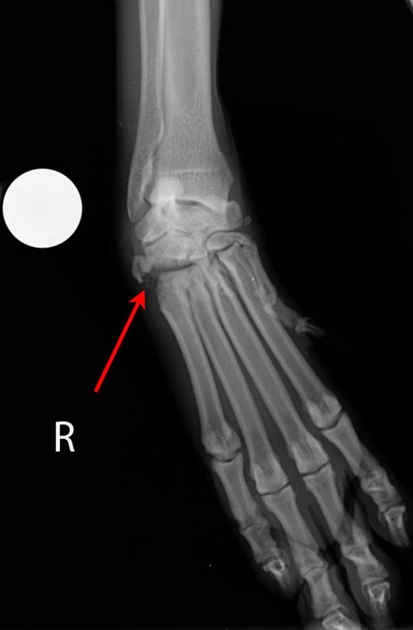
On the dorsopalmar varus stressed view (Figure 3), a marked widening of the lateral aspect of the carpometacarpal joint is present.

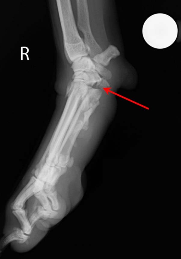
On the valgus stressed view (Figure 4), the intra-articular space of both the intercarpal and carpometacarpal joints appear to be wider than expected, suggestive of an injury involving the medial collateral ligament complex.

On the hyperextended mediolateral projection (Figure 5), a suspicion of palmar carpal instability exists. The fracture fragment arising from the proximal aspect of the fifth metacarpal bone can be appreciated (arrow).
The clinical and radiographic findings are suggestive of palmar and medial collateral ligament rupture, with a concurrent fracture of the proximal aspect of the fifth metacarpal bone.
Question
How would you manage the case?
Answer
Management options include:
- external coaptation
- surgical repair of the damaged ligament and stabilisation of the metacarpal fractures, possibly augmented by an external fixator
- arthrodesis
In cases where the palmar ligaments are damaged, external coaptation is not recommended, as it is associated with a poor prognosis and a high rate of bandage-related complications.
Surgical repair of the carpal collateral ligaments and metacarpal fractures can be attempted; however, lesions of the palmar ligament are associated with a high rate of implant failure.
Arthrodesis can either involve the intercarpal joints and the carpometacarpal joint only (partial carpal arthrodesis), or include the antebrachiocarpal joint (pancarpal arthrodesis). Partial carpal arthrodesis can only be performed when the surgeon is confident of an intact antebrachiocarpal joint.
In this case, pancarpal arthrodesis was considered a more predictable option for the management of the injuries.

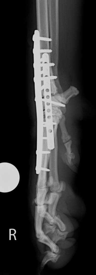
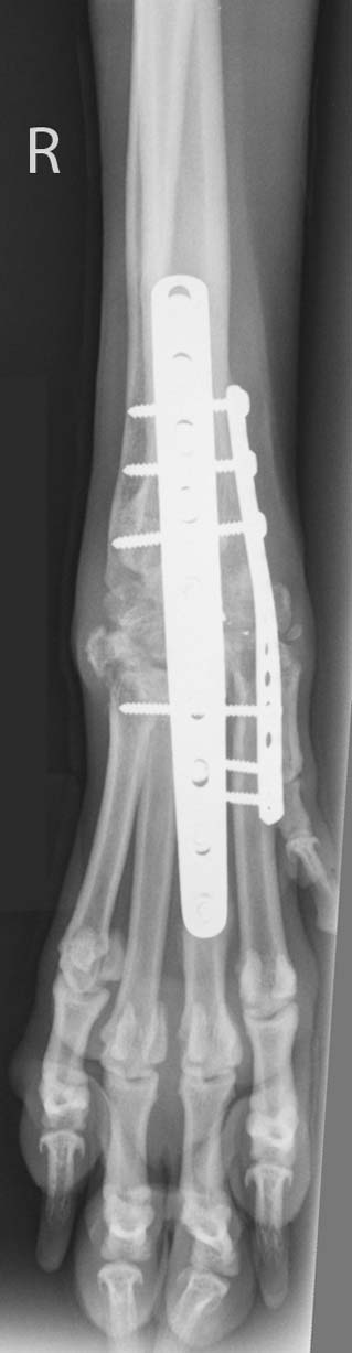
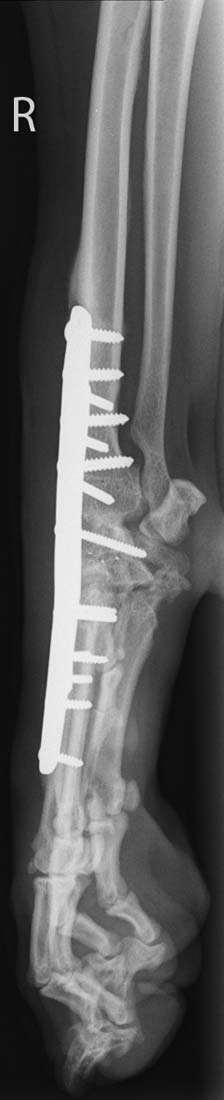
A craniomedial approach of the carpus was performed and the cartilage was removed from all joint levels using a high-speed burr. The intra-articular spaces were packed with demineralised bone matrix. A 3.5mm/2.7mm hybrid plate was applied dorsally, while a 2.4mm/2.7mm cuttable plate was secured on the medial aspect of the carpus. After closure, postoperative radiographs were performed (Figures 6 and 7). A padded dressing was applied. The dressing was replaced daily for four days. The patient was discharged on oral meloxicam and a seven-day course of cephalexin.
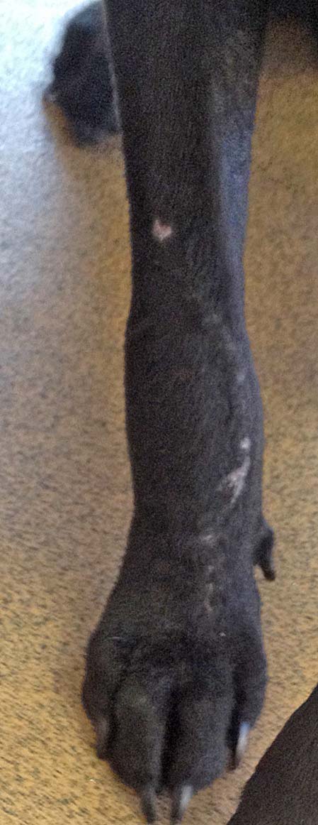
The owner was pleased to report a fast and progressive improvement of leg function. After three weeks, a mild lameness and mild carpal swelling were present (Figure 8). Radiographs obtained after eight weeks demonstrated excellent progression of arthrodesis and no evidence of implant complications (Figures 9 and 10).