5 Oct 2015
Christine Heinrich
Job Title
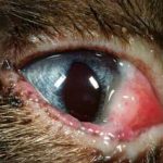
Figure 1. Non-specific conjunctivitis in a feline patient. Without other systemic or ocular signs, FHV-1, Chlamydophila felis and Mycoplasma species infection have to be considered. IMAGE: ©Christine Heinrich, Eye Vet Clinic.
In part one of this article, I outlined how primary infectious conjunctivitis was rare in dogs and that most canine patients present with conjunctivitis suffered from keratoconjunctivitis sicca (KCS) or adnexal anomalies.

In cats, the situation is opposite to dogs, in that primary infectious conjunctivitis is very common and conditions such as KCS and adnexal anomalies are rare. Unfortunately, in many cases it is difficult to distinguish the underlying infectious cause purely on clinical presentation and symptomatic treatment or laboratory investigations may have to be employed to reach a diagnosis. Specifically, infection with feline herpesvirus-1 (FHV-1), Chlamydophila felis and Mycoplasma felis may present identically (Figure 1) while the conjunctivitis in feline calicivirus (FeCV) infection has a more distinct appearance.
FHV-1 is the most common pathogen that will cause conjunctivitis in the cat. FHV-1 is ubiquitous in cat populations worldwide and can present as conjunctivitis alone or with keratitis and upper respiratory tract (URT) signs. In addition, a number of other corneal conditions, which will show concurrent conjunctivitis – including KCS, eosinophilic keratitis, corneal sequestration, superficial chronic corneal epithelial defects (SCCEDs) and chronic non-ulcerative keratitis – have been linked to FHV1 (Stiles, 2013).
FHV-1 is a DNA alpha-herpesvirus, which causes damage to the epithelial cells in which it replicates. FHV-1 is the cause of feline infectious rhinotracheitis and infection occurs by droplet spread nasally, orally and conjunctivally. Infected cats shed virus for one to three weeks following infection. Healthy individuals show replication of FHV-1 particles in the epithelium of the nose, throat, conjunctiva and cornea. The condition is often complicated by secondary bacterial infection.
Approximately 80% of all infected cats will become chronic FHV-1 carriers and the virus becomes latent in neurones of the trigeminal ganglion and associated nerve fibres as well as conjunctival cells and keratocytes. In approximately half of those carriers, the virus can be reactivated depending on a number of stress factors.
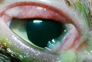
Clinical signs of FHV1 infection depend on the age and the immune status of the affected individual. Primary FHV-1 infection of an immune-competent adult cat often only results in a self-limiting mild URT infection and conjunctivitis. In these patients, clinical signs equal those of cats affected with Chlamydophila species or Mycoplasma infection (Figure 2a). If corneal ulceration occurs concurrently with conjunctivitis, it is considered pathognomonic for FHV infection as ulcerative keratitis is not believed to be a sign of either Chlamydophila species or Mycoplasma infection (Figure 2b). In some patients, however, corneal lesions are restricted to microdendritic ulcers, which do not show up with fluorescein stain, and only the use of rose bengal stain will allow identification of corneal involvement.
Treatment of conjunctivitis due to FHV-1 infection is usually limited to supportive management. This might purely involve the use of a lubricating eye gel for pets. Secondary bacterial infection is often suspected if the ocular discharge is purulent, but this is not necessarily the case as a neutrophilic infiltrate can be caused by the viral infection alone. However, the use of a suitable antibiotic, such as fusidic acid or chloramphenicol, may be indicated. Antiviral medications, both topical or systemic, are usually reserved for cases where severe respiratory tract signs of ulcerative keratitis occur. It must also be kept in mind antiviral drugs are not able to eradicate the virus from the host, but purely reduce the viral load and thus ameliorate clinical signs and virus shedding.
Diagnosis of FHV-1 infection can be confirmed by DNA testing. However, while being highly sensitive, the test is not necessarily giving conclusive evidence FHV-1 infection is the cause of the conjunctivitis – keep in mind that reactivation of latent virus and intermittent virus shedding are highly common even in healthy cats.
Feline calicivirus has been documented as a neglected cause of conjunctivitis. However, the serous conjunctivitis present in the affected cats was generally associated with systemic signs of URT infection and all affected cats were found to suffer from oral ulceration as a pathognomonic finding. Furthermore, unlike in conjunctivitis due to FHV-1, C felis and Mycoplasma infection, extensive ulceration of the conjunctiva in affected patients was documented while corneal ulceration was not a feature of this infection.

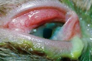
C felis is a Gram-negative bacterium that preferentially infects the conjunctiva, but also the mucosal membranes of the respirator and urogenital tract of cats. Infection occurs through droplet exchange in close contact, but the organism can also survive for a few days in the environment. Clinical signs are mostly limited to conjunctivitis, and the respiratory or urogenital signs are usually so subtle they are missed (Figure 3). Diagnosis of the disease is made with PCR testing, which is highly sensitive. Previously, diagnosis was often attempted by cytological diagnosis with the detection of cytoplasmic inclusion bodies. However, this technique is not very successful in chronic cases and PCR testing is preferred.
Conjunctivitis due to C felis infection readily improves on topical treatment with chloramphenicol ointment, but there is a tendency for clinical signs to recur. This is due to the fact the organism can lie dormant in the urogenital tract, which can be reactivated and reinfect the conjunctiva once topical treatment has been stopped. C felis is sensitive to treatment with doxycycline, potentiated amoxicillin, azithromycin and fluoroquinolones. A 28-day course of doxycycline 10mg/kg is reported to be most efficient at clearing the systemic infection, but care has to be taken to administer the drug with a bolus of water due to its risk to cause oesophagitis and strictures. The use of potentiated amoxicillin is recommended in kittens with C felis conjunctivitis to avoid the potential side effects of tetracyclines.
Recurring infection with C felis, even after appropriate treatment courses, can also be caused by contact with clinically normal individuals that shed the virus and act as a “silent reservoir”. All in-contact cats must therefore be treated to allow successful elimination of the disease in a household or in close proximity (Gruffydd-Jones et al, 2009). Accordingly, on occasions, cats belonging to neighbours may need to be included in the treatment. The use of Chlamydophila vaccines can be considered for high-risk cats and shelter situations, but the efficacy of such vaccination to entirely prevent clinical signs is not certain.
M felis, Mycoplasma gatae and Mycoplasma arginine have been implicated in the pathogenesis of conjunctivitis and, while many ophthalmologists have treated this organism as an incidental finding or opportunist one that occurs together with other pathogens, reports have made a firm link between the detection of the organism on PCR testing and conjunctivitis (Low et al, 2007; Hartmann et al, 2010). The organism can also cause lower respiratory tract disease in immunocompromised patients. Fortunately, most Mycoplasma species are sensitive to the same drugs used to treat Chlamydophila species.
A number of other bacterial agents, such as Bordetella bronchiseptica, may also have to be considered in cases of persistent feline conjunctivitis. However, such infections are usually only diagnosed after symptomatic treatment or testing for FHV-1, Chlamydophila and Mycoplasma have proven unfruitful. The organisms may be identified by bacterial culture and managed with appropriate antibacterial therapy.
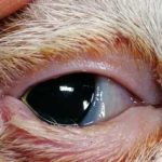
Conjunctivitis in cats can, of course, as in dogs, be caused by adnexal anomalies such as entropion. The latter is rare in cats, but if it occurs, it is often misdiagnosed as conjunctivitis (Figure 4). Eyelid colobomas are also rare, but again are likely to present as conjunctivitis.
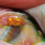
A rare condition reported in cats is eosinophilic conjunctivitis. This form of proliferative conjunctival inflammation is caused by infiltration of the conjunctiva – preferentially at the lateral lower lid – with eosinophils.
Clinically, the condition manifests with lid swelling, depigmentation and a caseous, “cottage cheese-like” exudate (Figure 5). Diagnosis is made on cytological examination of a scrape of the affected area, which can be obtained under local anaesthetic.
Many ophthalmologists believe this condition to be an early stage of feline eosinophilic keratoconjunctivitis (FEK), where similar lesions on the lids extend on to the cornea (Figure 6). The cause of the condition is not known and, while a link has been made between FEK and FHV-1 infection, this has not been proven for cases of pure feline eosinophilic conjunctivitis (Allgoewer et al, 2001). The condition responds to the use of topical steroids or ciclosporin and systemic treatment with megestrol-acetate, but must be restricted to severe cases given the potential side effects of this drug.
Also uncommonly seen is lipogranulomatous conjunctivitis. In this condition, lipid granulomas originating from the meibomian glands are visible as creamy, pale nodules underneath the conjunctiva near the lid margin. The aetiology of the condition is poorly understood, but an actinic effect on the meibomian glands, with subsequent “leakage” of sebaceous material into the subconjunctival tissue is believed to initiate the granulomatous inflammatory response, which is mostly seen in cats with little periocular pigmentation.
Treatment by curettage or surgical excision of the affected tarsal tissue is reported successful in managing the condition should it be associated with ocular irritation (Read and Lucas, 2001).
Given several infectious agents can cause a very non-specific conjunctivitis in cats, one might think this would warrant the immediate use of advanced laboratory investigations on diagnosis of feline conjunctivitis. However, not many clients would agree with such an expense on the first diagnosis of a relatively “minor” ailment. Obviously, the clinician should attempt to distinguish infection with FHV-1 (most likely to have concurrent corneal ulceration or systemic signs) from C felis and M felis infections.
The use of chloramphenicol eye ointment – which is efficient at managing the ocular signs of C felis and M felis infection – is an appropriate first-line treatment, and individuals with an adequate immune response are unlikely to present with a relapse of conjunctivitis once topical treatment is stopped. Equally, patients with FHV-1 infection, which only suffer from conjunctivitis as a visible sign, should recover on their own or with supportive topical antibacterial treatment.
Testing for FHV-1, C felis and Mycoplasma species with PCR testing as well as bacterial culture and sensitivity is usually restricted to those patients where the conjunctivitis fails to resolve on its own accord, is non-treatment responsive or recurrent.
The value of a positive FHV-1 PCR test is, as mentioned before, questionable, but the concurrent negative test for C felis or Mycoplasma species is highly significant. Equally, long courses for affected and in-contact cats to treat a C felis infection should not be prescribed without confirmation of the infection by PCR testing first.