10 Apr 2017
Negar Hamzianpour with the latest in the Case Notes series.
Negar Hamzianpour
Job Title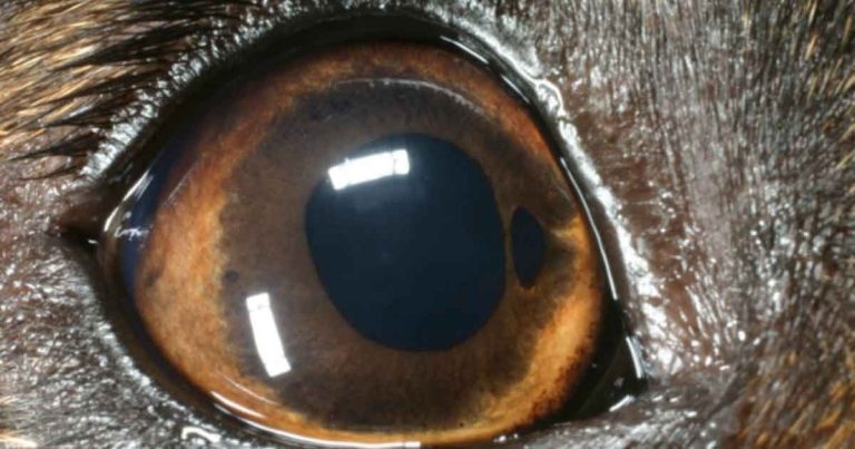
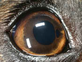
Figure 2. Iris coloboma in a three-year-old cross-breed dog.
Your first case of the morning is a routine vaccination for a nine-year-old female neutered toy poodle.
The owner raised concerns the dog was squinting outdoors and had dilated pupils.
A general physical exam revealed the dog was otherwise well. Ophthalmic examination confirms bilateral mydriasis (Figure 1), but that both eyes are visual. Further questioning reveals the patient has demonstrated no obvious visual deficits at home.
What is your differential diagnosis list for this dog, what further questions would you ask
the owner and what further testing could
you perform?
The differential diagnosis list for bilateral mydriasis in a visual dog includes:
It is important to obtain a full detailed history, and perform a general physical and neurological examination. In this case:
By looking back at your original differential diagnosis list, what is the most likely diagnosis and why?
Iris atrophy is the most likely diagnosis in this case. Iris atrophy is degeneration and thinning of the iris constrictor muscle. It can be primary or secondary to diseases, such as glaucoma or uveitis.
This case is an example of primary iris atrophy, also known as senile iris atrophy.
It is a spontaneous progressive thinning of the iris in middle-aged to older dogs of any breed, although it is more commonly seen in poodles and cocker spaniels. It is characterised by a scalloped, moth-eaten appearance of the pupillary margin.
Changes can be asymmetrical between the two eyes, cause dyscoria and lead to reduced PLRs. It is often difficult to clearly appreciate the PLRs in these cases, but, on close examination, there is at least a slight constriction of the iris with bright light. An important practical tip in general practice is to use a very bright focal light source.
In practice, few pointers can help you make this diagnosis. It is important to note vision is not affected by iris atrophy. Although, vision in bright light conditions may be slightly affected due to photophobia.
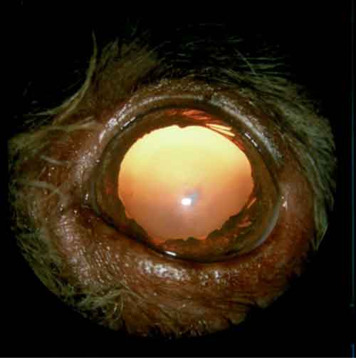
Retro-illumination, when light is reflected from the tapetal fundus through the areas of the affected iris, can assist in highlighting the thinning of the iris.
Finally, you could consider applying a drop of a topical prostaglandin analogue (for example, travoprost) to the eye. A normal side effect of this drug in dogs is miosis.
In cases of iris atrophy, due to degeneration of the iris constrictor muscle, miosis will be significantly reduced or not occur at all.
Although topical prostaglandin analogues are used in cases of canine glaucoma to lower intraocular pressure, this effect is not of concern in normotensive eyes. It is, however, important to note these drugs are contraindicated in cases of anterior lens luxation and should be used in caution in cases of anterior uveitis.
Another key differential is congenital iris hypoplasia, also known as an iris coloboma. This is due to incomplete iris development, rather than degeneration. This can be distinguished from iris atrophy as it is present from birth, is non-progressive and less diffuse than iris atrophy (Figure 2). Typically, this will present in younger animals.
What treatment is needed in this case and what can you advise the owner?
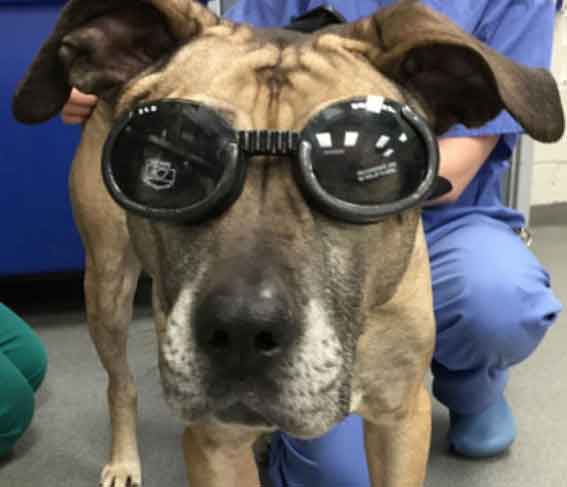
Iris atrophy is a relatively benign, but progressive, condition. In severe cases it can cause photophobia. The use of tinted dog goggles, if tolerated, outside on bright sunny days, can be suggested to owners to improve comfort (Figure 3).
In very severe cases the lens can subluxate due to concurrent atrophy of the ciliary processes and zonules. However, this is seen rarely and does not normally lead to secondary glaucoma.
Iris atrophy is a common condition seen in practice in middle-aged to older dogs. It is usually an incidental finding that can be diagnosed during the consultation by performing a complete history and physical examination. It rarely requires any further treatment or diagnostics.
The author would like to thank Mike Rhodes for his support and assistance with this article.