18 Nov 2019
Francesco Cian discusses the case of a four-month-old male mixed-breed dog with a history of anorexia, weight loss and diarrhoea in his latest Haematology Hub.
Francesco Cian
Job Title
Figure 1. The blood smear image.
The image in Figure 1 (Wright-Giemsa, 50×) and the data (Table 1) are from an ethylenediaminetetraacetic acid blood sample of a four-month-old male mixed-breed dog, seen by the referring veterinarian with a history of anorexia, weight loss and diarrhoea with dark, tarry faeces.
Based on the data and the blood smear image provided, what is the most likely explanation for the anaemia observed?
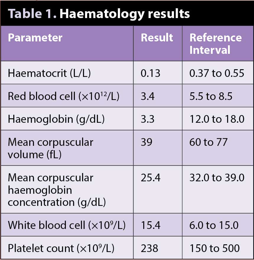
Automated blood cell count reveals severe anaemia, confirmed on blood smear examination, which shows a significant decrease in red blood cell density.
Most red blood cells appear microcytic (low mean corpuscular volume) and hypochromic (low mean corpuscular haemoglobin concentration) as a result of defective haemoglobin synthesis and red blood cell maturation, both caused by iron deficiency.
Hypochromasia can be appreciated on blood smear as increased central pallor visible in most red blood cells.
Iron deficiency anaemia occurs when either dietary iron intake does not meet the body’s requirement or chronic external blood loss is present. Inadequate intake is very unlikely in dogs fed commercial pet food or balanced home-cooked diets. Chronic external blood loss is much more common and often affects the gastrointestinal tract. Gastrointestinal haemorrhage can result from primary gastrointestinal disease (such as parasitism and neoplasm), ulcerogenic drugs (such as corticosteroids and NSAIDs) or may be secondary to systemic diseases (such as coagulation disorders, hepatic diseases and hypoadrenocorticism).
Iron deficiency anaemia develops after weeks to months of chronic or recurrent blood loss and is often regenerative in the first phases; with depletion of iron body stores, it then becomes non-regenerative, microcytic and hypochromic.
Diagnostic imaging is often warranted to further investigate iron deficiency anaemia, especially when gastrointestinal bleeding is suspected. Faecal occult blood test may also be performed; however, both false negative and false positive results may occur.
Iron status may be further investigated by measuring serum iron parameters and checking iron stores in the bone marrow.
Faecal analysis revealed the presence of large numbers of eggs of Ancylostoma caninum – a nematode also known as hookworm due to the hook-like mouthparts used to anchor to the lining of the intestinal wall.
This parasite is commonly found in most tropical and subtropical areas, although infections are also seen in cooler regions where the temperature is suboptimal. In those areas – including the UK – another hookworm called Uncinaria stenocephala prevails.
Hypochromic, microcytic anaemia due to gastrointestinal chronic blood loss may occur in puppies with heavy infestations and may be fatal.
Hookworms may also cause formation of characteristic cutaneous lesions by larvae penetrating the pads, interdigital skin, and skin in contact sites.