24 Sept 2018
Francesco Cian discusses the case of a 1.5-year-old female crossbreed dog imported from eastern Europe in his latest Cytology Corner.
Francesco Cian
Job Title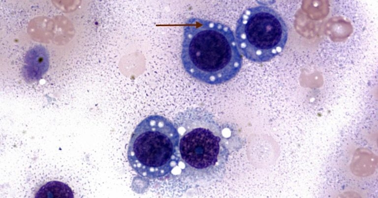
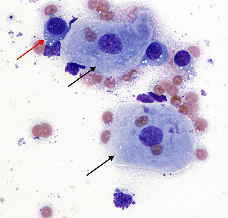
These two microscopic images (Wright-Giemsa stain 100× [Figure 1] to 50× [Figure 2]) are from a vaginal swab of a 1.5-year-old female crossbreed dog imported from eastern Europe. The dog had apparently been neutered and the vet suspects an ovarian remnant syndrome as a cause of the vaginal discharge.
What is your interpretation?
The submitted smear is moderately cellular with adequate preservation. The background is lightly basophilic with moderate numbers of red blood cells. A main population of discrete, round cells exists, together with a few superficial vaginal squamous epithelial cells (black arrows) and rare neutrophils.
The round cells (red arrows) have moderate amounts of deeply basophilic cytoplasm with defined borders – often containing clear, distinct, intracytoplasmic vacuoles – mostly arranged in a linear array along the inner surface of the cell membrane. Nuclei are round, centrally to paracentrally located, with granular chromatin and single, round nucleolus occasionally seen. Anisocytosis (cell size variation) and anisokaryosis (nuclear size variation) are mild. Rare mitotic figures (not seen in the images) are present.
 The aspirate harvested a main population of discrete round cells with distinctive cytologic features, compatible with transmissible venereal tumour (TVT) and further supported by the location of the process.
TVT is a histiocytic tumour of the external genitalia, described in the dog and other canines living in temperate climates. Preferred locations include external genitalia, and all the mucous membranes associated with sexual contact. TVT usually remains localised, but metastases to regional lymph nodes and other organs may occur in a small percentage of cases. TVT is primarily spread by coitus and free-roaming, sexually intact, mature dogs of any breed, age or sex are at greatest risk.
TVT and Tasmanian devil facial tumour disease are the only known naturally occurring clonally transmissible cancers that behave like an infectious parasitic neoplastic tissue graft. These neoplasms have overcome the limitations of existing within the single host and have gained the ability to spread between individuals, therefore surviving long after the original hosts have died.
In this specific case, a more detailed inspection of the vaginal canal confirmed the presence of a characteristic friable, cauliflower-like mass, oozing a serosanguineous fluid and likely the cause of the vaginal discharge. The tumour may arise deep in the prepuce or vagina and it may be difficult to see; this may lead to misdiagnosis if bleeding is confounded with oestrus, urethritis, cystitis or prostatitis.
The lesion underwent complete regression after four weekly doses of vincristine.