8 Jan 2018
Francesco Cian presents the latest from the Cytology Corner series, with pictures from an aspirate of a nodular lesion on the trunk from an adult male.
Francesco Cian
Job Title
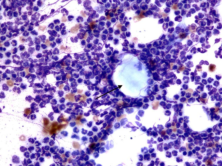
Pictures from an aspirate of a nodular lesion on the trunk from an adult male dog (Wright-Giemsa stain, 10× to 50×).
These pictures are from an aspirate of a nodular lesion on the trunk from an adult male dog (Wright-Giemsa stain, 10× to 50×).
The submitted smear is highly cellular with adequate preservation. The background is clear with occasional red blood cells and variable numbers of anucleated keratinocytes (black arrow).
Large numbers of segmented neutrophils are present, often forming groups of variable sizes and surrounding the keratinocytes. Occasional reactive macrophages with abundant foamy cytoplasm (not seen in these pictures) and rare cholesterol crystals are also noted.
Inflamed follicular cyst.
The presence of anucleated keratinocytes is a common finding observed in lesions of follicular origin. These include follicular cysts and follicular tumours – the former considered more likely in this case, given the absence of nucleated epithelial cells.
Follicular cysts are non-neoplastic, simple, sac-like structures with an epithelial lining. In dogs, they often appear as solitary, nodular lesions, mainly localised to the trunk and head. In selected breeds, such as German shepherd and Pekingese, they can be multiple. Classification of cysts depends on identification of the lining epithelium or the pre-existing structure from which the cyst arose.
Cysts of follicular origin can be categorised on histopathology, based on the level of the follicle from which they develop into:
The rupture of any type of keratinous cyst may result in a marked foreign body-type inflammatory reaction in the dermis, as observed here, with keratinocytes surrounded by inflammatory cells (such as neutrophils and macrophages).
The alcoholic fixing of slides can also cause the dissolution of the cholesterol present in the lipid envelope of the cells, with formation of characteristic cholesterol crystals represented by transparent, rectangular structures.