15 Jul 2019
Francesco Cian presents haematology slides from another case in the latest in his Veterinary Times series.
Francesco Cian
Job Title
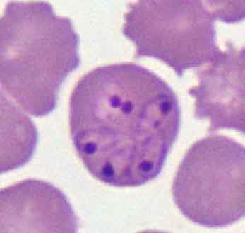
Figure 1. A blood smear from the dog.
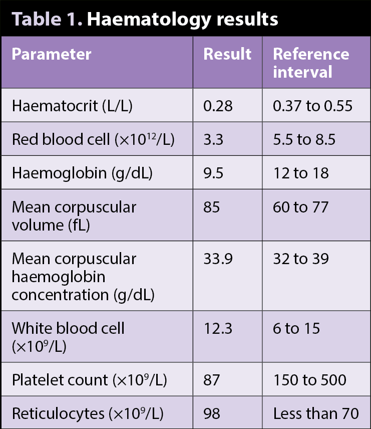
The images (Figures 1 and 2; Wright-Giemsa 50 to 100×) and data (Table 1) are from an ethylenediamine tetra-acetic acid (EDTA) blood sample of an adult, male dog seen by the referring veterinarian for having been lethargic and anorexic for a week. The dog had history of travelling to southern Europe.
The reported anaemia appears to be regenerative, given the increased number of reticulocytes provided by the analyser and the subjective increase in polychromatophils seen on the blood smear. Regenerative anaemia may be caused by blood loss and/or haemolysis. The latter is more likely in this case, especially given the presence of intracellular parasites within some of the red blood cells (red arrows).
These appear as poorly staining, teardrop-shaped structures with an eccentric, deeply basophilic nucleus. Their morphology is compatible with Babesia species, which is a haemoprotozoan organism transmitted by various types of tick, and is often the cause of haemolytic anaemia and thrombocytopenia in domestic animals. Babesia species varies in appearance, and both large (2μm to 5μm) and small (1μm to 3μm) forms have been described. Large forms include Babesia canis – as seen in this case.
Blood smear examination is considered highly specific, since the presence of the parasite is indicative of infection. However, false negative results are common and PCR testing is recommended in all those suspected cases where parasites are not visible on blood smear examination. PCR also has the advantage of being able to further classify the parasite, as the pathogenicity may vary depending on the species.
Red blood cells with frequent sharp spicules are referred to as echinocytes, burr cells or, more simply, crenated erythrocytes (blue arrow). They are often an artefact that results from excess EDTA, improper smear preparation or prolonged sample storage before blood film preparation. In dogs, echinocytes may also be observed in snake envenomation, uraemia, blood transfusions, underlying neoplasia, and rarely in genetic disorders affecting the red blood cells, including pyruvate kinase deficiency.