11 Feb 2019
Francesco Cian continues his Haematology Hub series by discussing a case of a cat with a clinical history of pyrexia, soft faeces, inappetence and intermittent vomiting.
Francesco Cian
Job Title
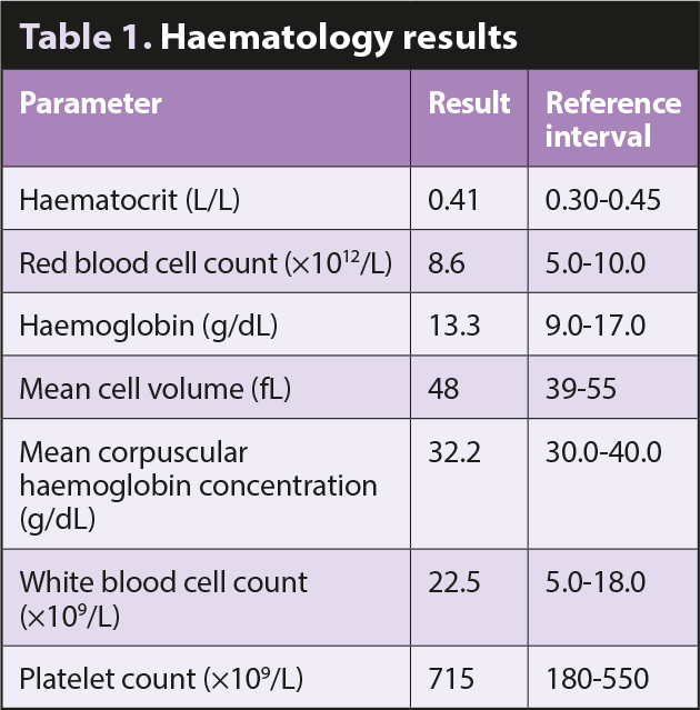
Figure 1. Wright-Giemsa stain 50×.
The image (Figure 1, Wright-Giemsa 50×) and data are from an ethylenediamine tetra-acetic acid (EDTA) blood sample of an adult, domestic short-haired cat with a clinical history of pyrexia, soft faeces, inappetence and intermittent vomiting.
Based on the data and blood smear picture provided, try to answer the following questions:
Blood smear examination confirmed the presence of mild leukocytosis; the majority of the leukocytes observed are segmented eosinophils (65%), which can be easily recognised on microscopy due to the presence of characteristic intracytoplasmic, rod-shaped, pink/orange granules (red arrows).
Eosinophilia in cats has been associated with parasitic and infectious diseases, hypersensitivity disorders, eosinophilic infiltrative diseases, and underlying neoplasia. In particular, paraneoplastic eosinophilia has been reported in association with mammary tumours, leiomyosarcoma, T-cell lymphoma, fibrosarcoma, mast cell tumour and urinary bladder tumours.
Imaging studies on this case identified the presence of an intestinal thickening and mesenteric lymph node enlargement, which was confirmed to be a T-cell lymphoma by cytology and flow cytometry. Eosinophil chemotaxis may be a response to the production of interleukin-5 by neoplastic lymphoid cells.
Thrombocytosis is a relatively common finding and has been reported in approximately 5% of feline patients from a busy UK small animal hospital (Rizzo et al, 2007).
Platelet numbers (blue arrow) can arise due to several conditions, including physiological thrombocytosis, reactive thrombocytosis or essential thrombocythaemia – the latter only rarely described.
Physiological thrombocytosis is very common and is the result of the mobilisation of platelets from body stores in response to exercise or excitement/stress. Reactive thrombocytosis occurs as a rebound response of the bone marrow to several conditions, including inflammation and neoplasia, and is, therefore, considered more likely in this case.
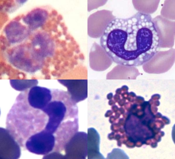
Eosinophils may appear very different depending on the animal species and sometimes even the breed (Figure 2).
In dogs (top left), eosinophils have round, orange granules that may appear clear in greyhounds (and most sighthounds), hence the name ”grey eosinophils” (top right).
In cats (bottom left), eosinophils have small, rod-shaped orange to pink granules that fill all the cytoplasm, whereas those of horses (bottom right) have very large, globular, red/orange granules, giving the cell a raspberry-like appearance.