23 Oct 2015
Hany Elsheikha
Job Title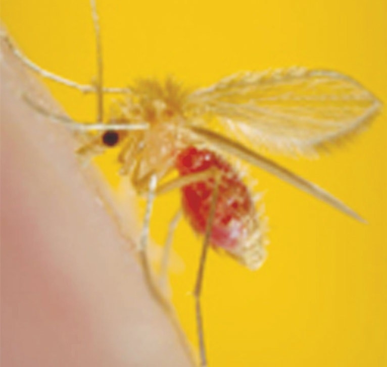
Figure 2. The insect vector of leishmaniosis, sandfly Phlebotomus papatasi (Diptera: Psychodidae: Phlebotominae) feeding on a mammalian host. The female sandfly carries the Leishmania organisms in its salivary glands and injects them into the host as it feeds. Note the red colour of the fly’s abdomen, which is full of the ingested host’s blood. Image: ©CDC/Public Health Image Library.
Canine leishmaniosis (CanL) is an important emerging vector-borne disease caused by the protozoan parasite Leishmania infantum. Its increasing prevalence in dogs in Europe has been a serious concern to pet owners, veterinarians and public health officials, and has created a growing challenge that requires a one health approach to counter the spread of the infection to new, non-endemic regions.
The majority of dogs in endemic areas remain sub-clinically infected because the clinical disease affects only a proportion of infected dogs. A combination of genetic and immunological mechanisms and, possibly, other as yet unknown factors can cause such variability in the clinical response to Leishmania infection.
Despite the lack of a gold-standard test, diagnosis of CanL can be established based on a combination of clinical signs compatible with the disease plus serology (demonstration of specific immune responses against Leishmania), and the detection of the parasite DNA in a dog’s tissues as determined by PCR. Correct and early diagnosis is essential for timely institution of treatment and for minimising transmission of Leishmania from dogs to vectors.

Leishmaniosis is a vector-borne protozoan zoonotic disease caused by Leishmania species, which are transmitted mainly via the bite of the phlebotomine sandflies belonging to the genera Phlebotomus (old world; Africa, Asia, Europe) or Lutzomyia (new world; the Americas). This disease has been reported from about 98 countries (Alvar et al, 2012).
Canine leishmaniosis (CanL) is caused by Leishmania infantum (L chagasi); the only Leishmania species reported in both old and new worlds, and can potentially cause fatal disease in humans and dogs.
Historically considered to be a neglected tropical and subtropical disease, CanL has become endemic in many temperate regions with at least 2.5 million dogs reported to be infected in south-western Europe. This makes CanL a very important disease from both veterinary and public health perspectives, especially with the increased travel of pets across Europe and the growing number of imported dogs.
There is a clear need for implementation of effective prophylactic measures to prevent the disease becoming established in the UK. Veterinary professionals must remain up-to-date with developments in the clinical evaluation, diagnosis and management of CanL to provide the best advice to their clients taking dogs to mainland Europe and to provide better management of clinical cases, which are expected to increase in the near future.
The life cycle of the protozoan Leishmania is indirect (it requires a vector host plus the main mammalian host) and alternates between two main developmental forms (Figure 1): the amastigote and the promastigote. In the mammalian host, Leishmania species occurs as amastigotes (~2μm to 5μm in diameter) solely within mononuclear phagocytes in the skin, bone marrow and visceral organs.

In the gut of dipteran sandflies (genus Phlebotomus), the sole known vector in Europe, Leishmania species occurs as flagellated, extracellular promastigotes (~15μm to 30μm in length). The acquisition of L infantum infection occurs through the bite of the haematophagous sandflies (Figure 2) when a feeding sandfly deposits metacyclic promastigotes into the dermis of the mammalian host. After local multiplication of the parasites in dendritic cells and macrophages in the skin, dissemination occurs via the lymphatic system and blood. Thus, Leishmania parasites can be found in the skin, lymph nodes, spleen, liver, bone marrow and other organs.
Dogs are the primary reservoir for L infantum (Quinnell and Courtenay, 2009), due to their high susceptibility to the infection and their ability to transmit the parasite to the arthropod vector, especially by dogs with evident clinical disease. Dogs also represent a major source of human infection.
Other, less frequent, forms of non-vectorial transmission between dogs can occur via transfused blood products from infected donors, and transplacental and venereal transmission. Human outbreaks in Spain (Arce et al, 2013) that were not accompanied by any increase in the prevalence of CanL have led to speculation other animals, such as wild rabbits, might be able to transmit Leishmania to sandflies, and thus can act as reservoirs (Jiménez et al, 2014).
Other animal species, including golden jackal, Iberian lynx, mustelids, red fox, wild cat, and wolf, have also been diagnosed with Leishmania infection (Millan et al, 2014), but their role as sources of infection (infectiousness to sandflies, persistence of infection) remains unknown. Interestingly, L infantum RNA has been detected in fleas and ticks collected from infected dogs. However, the competence of ticks or fleas as a vector and proof of transmission has not been confirmed.

In Europe, three Leishmania species have been reported – L infantum, L tropica and L donovani. L infantum is the predominant species causing CanL, and the visceral leishmaniasis (VL) and cutaneous leishmaniasis (CL) forms in humans. L tropica and L donovani occasionally can cause CL and VL in humans. The seroprevalence in dogs ranges from 5% to 30%, and can reach 90% depending on the geographic region. CanL is endemic in Europe in Albania, Croatia, Cyprus, Greece, France, Italy, Malta, Portugal and Spain.
CanL has shown increasing trends in both the prevalence in traditional endemic regions and the expansion towards new, non-endemic locations, most probably driven by the increased mobility of dogs, international tourism and climatic changes. The latter can largely influence the geographic distribution (Figure 3), seasonal abundance and vectorial capacity of sandflies, which affects the distribution of leishmaniosis.
The main risks in endemic areas are related to vector exposure and abundance of reservoir dog hosts including hunting dogs, stray dogs, and those living outdoors or adopted from animal shelters.
About 693 cases of leishmaniosis have been reported in dogs imported and/or resident in the UK (Nash, 1993; Manser, 2004).
Another study reported 257 clinical cases of CanL between 2005 and 2007 (Shaw et al, 2009). These cases had spent at least six months in southern Europe, were rescued from rehoming centres in the country of origin, or entered the UK with confirmed infection. The spectrum of clinicopathological signs in some dogs was similar to that reported in endemic countries (Shaw et al, 2008, 2009).
The pet travel scheme, introduced in 2000, has relaxed the restriction on the movement of dogs from the EU continental endemic countries to the UK (Shaw et al, 2003; Manser, 2004), which might have facilitated the introduction of exotic infections. The number of dogs travelling to the UK has increased, with about 411,582 dogs recorded between 2000 and 2008 (Mencke, 2011). Potentially, a significant reservoir of dogs is infected, which is expected to increase due to the importation of infected dogs and the progression of global warming and which, with the lack of local prophylactic measures, may allow competent vectors of this exotic disease to become established in the UK.

Despite the lack of pathognomonic signs, dogs with leishmaniosis often present with lymphadenomegaly, dermatitis (Figure 4), alopecia, onychogryphosis, lameness, anorexia, weight loss, conjunctivitis, epistaxis, anaemia and renal dysfunction.
Atypical manifestations include mucosal lesions, osteolytic and osteoproliferative lesions, chronic colitis, splenomegaly and hepatomegaly, and disorders of the cardiovascular, respiratory, nervous and musculoskeletal systems.
A system of four clinical stages from mild to severe disease based on clinical signs, clinicopathological/laboratory findings and level of serum anti-Leishmania antibodies has been proposed (Table 1). This system should be taken into account when deciding which treatment is best for the individual patients and when making clinical judgement based on the prognosis for each stage (adapted from Solano-Gallego et al, 2009).
CanL is a disease in which infection does not parallel clinical illness, and this is reflected in the high prevalence of subclinical infection among endemic dog populations.
The incubation period (time from infection until the appearance of disease) of L infantum varies from months to years and the clinical aspects of infections range from a total absence of signs to a severe systemic and even fatal disease. This variability may result in diagnostic uncertainty, therapeutic delays, and the misuse or overuse of anti-Leishmania drugs.
Two main mechanisms have been postulated to explain the variability in the clinical response of dogs to infection.

A complex genetic background is suspected of influencing susceptibility of dogs to Leishmania infection. There is evidence of resistance in certain dog breeds (such as the Ibizan hound) that rarely develop the clinical disease, probably due to the presence of a protective cellular immunity.
On the other hand, several dog breeds, including German shepherd, cocker spaniel, Rottweiler and boxer, are more susceptible to disease. No sex or age dependent risks have been described. However, the disease is commonly seen in dogs younger than three and older than eight, and more probably associated with immunocompromising conditions. Some studies have shown disease is more common in male dogs, although this is not a consistent finding.
Host defence has been shown to rely mainly on the activation of a protective type-one T-helper cell-mediated immune response. Production of tumour necrosis factor-α (TNF-α) and interferon-γ (INF-γ) activates macrophages to destroy the amastigotes via nitric oxide-mediated mechanisms.
In contrast, the progression to disease is associated with a reduced T-cell mediated immunity and a marked type-two humoral response. Indeed, humoral immune response does not seem to be protective against infections caused by Leishmania. Besides being non-protective, anti-Leishmania antibodies are responsible for hyperglobulinaemia and immune complex-mediated lesions in various body organs, such as the kidney and eye, which can lead to variable clinical signs and non-specific clinicopathological findings.
An inflammatory response, high levels of serum anti-Leishmania antibodies and reactive oxygen intermediate production (oxidative stress), and decrease in CD4+ T-cell count with loss of antigen-specific proliferation are more evident in dogs exhibiting overt clinical signs, which correlate with high parasite burden, increasing the risk of sandfly infection. In endemic areas, the presence of high antibody levels does not necessarily imply the illness is due to Leishmania infection, because subclinical infection is common. Also, although clinical signs together with relevant epidemiological information can suggest a putative diagnosis, further work-up is necessary to confirm clinical leishmaniosis by other diagnostic methods, such as cytology, histopathology and PCR. A clinical algorithm for diagnosing CanL is provided (Figure 5).
Clinical signs are only observed in a proportion of infected dogs, which can lead to the apparently healthy infected dogs being misdiagnosed and left untreated, leading to an increase in the reservoir of infection for sandflies.
For these reasons and because Leishmania infection in dogs shares physical signs and clinicopathological abnormalities with other canine diseases, laboratorial confirmation of infection is necessary.
Direct diagnosis is possible via the detection of the amastigote stage of the parasite in Giemsa or Diff-Quik stained smears obtained from superficial lymph nodes or bone marrow aspirates of clinically affected dogs or after culture of samples to allow the development of promastigotes in vitro (Maia and Campino, 2008).
Parasite culture requires technical expertise and special media, and one month of incubation time might be needed. Staining is less sensitive in detecting the parasite in skin biopsies, but immunohistochemistry techniques, such as immunoperoxidase staining, can enhance the sensitivity of detection.

Serologic detection of specific anti-Leishmania antibodies is the most useful and most routinely performed diagnostic test for CanL (Solano-Gallego et al, 2009). It is relatively non-invasive and permits the detection of a specific antibody response in dogs at around six to eight weeks after an initial infection. In subclinical infections this period may extend to years.
Several tests are available for serodiagnosis of CanL, including three ELISA tests, three immunochromatographic tests (ICT), direct agglutination test, western immunoblot and immunofluorescence antibody test.
These tests vary in sensitivity and specificity according to the defined cut-offs and, although they verify the presence of antibody, they do not prove or rule out active infection. Also, these assays cannot differentiate between infected and vaccinated dogs. Further, these assays may give false-positive reactions with sera of dogs imported from endemic areas that have been vaccinated against Leishmania and in dogs infected by Trypanosoma cruzi, another protozoan that infects dogs in the Americas.
However, the recombinant K39 dipstick immunoassay does not cross-react with T cruzi or Babesia. ICT devices are particularly useful because they are simpler to perform compared to other traditional antibody tests and they produce a result in minutes (Solano-Gallego et al, 2009; Kalayou et al, 2011).
A 2014 study reported very high sensitivity and specificity using dried blood spot samples collected and stored on a blood collection device designed for storage and transport at ambient temperature to detect antibodies against L infantum in dogs using a commercial ICT assay compared to ICT results using thawed plasma (Rosypal et al, 2014).
PCR techniques are the most sensitive for diagnosis of infection by detecting Leishmania DNA in dog tissues, with aspirates of lymph node, bone marrow, or spleen, skin biopsies, and conjunctival swabs preferable (higher diagnostic sensitivity) over blood, buffy coat or urine (Maia et al, 2009; Solano-Gallego et al, 2011).
PCRs, targeting repetitive sequences, have been very sensitive compared with in vitro cultivation and have little risk of bacterial contamination.
The diagnostic sensitivity however, depends on the quality of the clinical samples. Lymph node aspirates, especially from animals with a lymphadenopathy, are the most convenient while bone marrow sampling is more invasive, but may be indicated for cases such as suspect asymptomatic animals.
PCR assays are available at some veterinary diagnostic laboratories and can be used to confirm infection and to monitor the efficacy of treatment.
A distinctive characteristic of L infantum is its capacity to enter and replicate within macrophages.
The typical histopathological finding in the affected tissues is an inflammatory reaction associated with macrophages with the presence of a variable number of Leishmania amastigotes within macrophages. Immunohistochemistry can be used to detect Leishmania when the parasite cannot be seen with routine histopathology. Lymphoplasmacytic inflammation is also common in dogs with leishmaniosis.
Histopathological lesions have been found more commonly in spleen, lymph nodes, bone marrow, liver, gastrointestinal tract and skin – organs that seem to have abundant mononuclear phagocytic cells. Nasal cavity and eyes may develop similar inflammatory patterns. A reactive lymphoid hyperplasia may be found in lymphoid organs including spleen and lymph nodes.
Although the laboratory findings may be variable, there are many findings such as a normocytic, normochromic, non-regenerative anaemia and, less frequently, thrombocytopenia, plasma protein changes with hyperglobulinaemia and hypoalbuminaemia, proteinuria, and a variable azotaemia with an increase in the UPC ratio due to renal dysfunction.
Hence, renal function should be evaluated in all infected dogs. These abnormalities should raise the index of suspicion for CanL in any at-risk dog.
Ultrasonography in dogs with visceral leishmaniosis can reveal enlargement of abdominal lymph nodes, spleen and liver.
Radiographic examination of long bones of affected dogs with bone involvement can show periosteal proliferation, changes in the intramedullary opacity, and/or cortical and medullary destruction.
In dogs with VL differential diagnosis (DD) includes babesiosis, histoplasmosis, brucellosis, systemic lupus erythematosus and canine monocytic ehrlichiosis. In dogs with skin lesions, DD includes mange (demodectic or sarcoptic), pyoderma, and fungal (Malassezia) dermatitis.
L infantum is the causative agent of human and canine leishmaniosis in Europe. Infection spreads primarily via the bites of infected sandflies. Infection does not always lead to clinical illness and dogs with a Th2 response tend to develop severe, progressive disease, while dogs with a Th1 immune response tend to develop self-limiting disease.
No gold standard diagnostic methods exist; however, direct microscopy, culture, histopathological examination of clinical samples and serological and molecular methods are available and can be used to aid diagnosis.
The importance of CanL in Europe is growing due to the effects of climatic and environmental changes on the sandfly vector of the disease, the increased northwards expansion of the infection from southern European endemic regions, increasing travel of humans with their dogs, inadequate health control of stray and sheltered dogs especially in endemic areas – which increases the pool of reservoirs – the difficulty associated with the detection of CanL due to the long and variable incubation period and polymorphic clinical course of the disease, and the threat of exotic or new Leishmania species.
These challenges require developing more effective preventive and therapeutic measures to counter the spread of this serious disease.