12 Apr 2021
Francesco Cian presents the case of a one-year-old, female, mixed breed dog in the latest in his Diagnostic Dilemmas series.
Francesco Cian
Job Title
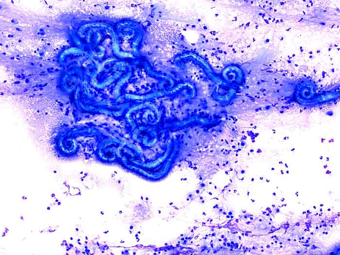
Figure 1. Peripheral blood, dog (Wright-Giemsa 50×).
The blood smear photomicrograph (Wright-Giemsa 50x; Figure 1) and the haematology data (Table 1) are from an ethylenediamine tetra-acetic acid blood sample of a one-year-old, female, mixed breed dog presented to a local veterinary clinic with acute respiratory symptoms (dyspnoea, coughing), loss of appetite and exercise intolerance.
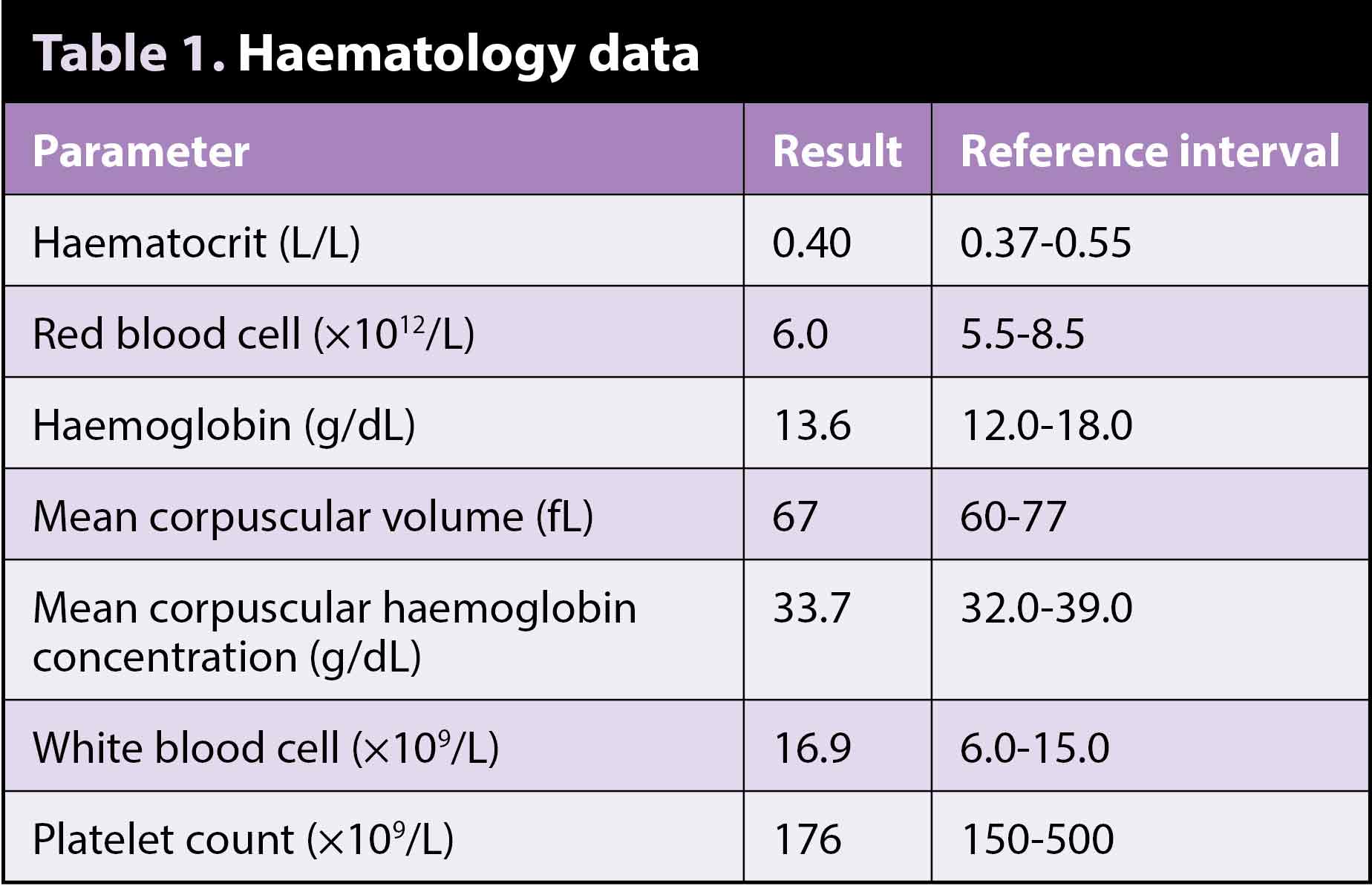
Based on the data and the blood smear picture provided, try to answer the following questions.
Examination of the blood smear confirmed the presence of mild leukocytosis. Admixed with neutrophils, there were increased numbers of eosinophils (orange arrows) and basophils (purple arrow), confirming eosinophilia and basophilia, respectively.
Eosinophils are polymorphonuclear cells slightly larger than a neutrophil, characterised by typical intracytoplasmic orange granules, whereas basophils contain granules of similar size and shape, but are purplish in colour.
Eosinophilia is a relatively common finding in the canine species. It often accompanies hypersensitivity and allergic processes, but can also be seen in association with parasitic infections, it can be paraneoplastic (for example, mast cell tumour, lymphoma) or idiopathic and it can also accompany hypoadrenocorticism.
Chronic eosinophilic leukaemia may also occur, but is an extremely rare event and is usually diagnosed by exclusion of other causes of persistent eosinophilia. Basophilia is a much rarer finding. It is often observed in association with eosinophilia and shares similar causes.
Other more specific causes for selective basophilia include essential thrombocythemia (extremely rare chronic myeloproliferative neoplasm characterised by marked increased number of platelets in the blood) and chronic basophilic leukaemia.
Given the clinical history of acute respiratory symptoms and the concurrent eosinophilia/basophilia, the veterinarian requested the antigenic test for Angiostrongylus vasorum to be run from the serum of this patient. This was positive, confirming the diagnosis of lungworm infection.
Alternative ways to reach a diagnosis of A vasorum (but not performed in this case) include:
Since coagulopathies often accompany cardiopulmonary disorders caused by this lungworm, coagulation testing (for example, platelets, prothrombin time, partial thromboplastin time, D-dimers) are also recommended.
A vasorum (also known as French heartworm) is a nematode found in most of Europe and parts of North America; it is endemic throughout most of the UK.
Adults are commonly observed in the right side of the heart and pulmonary arteries of dogs. Eggs hatch quickly and first-stage larvae migrate through the capillary beds into the bronchioles, bronchi and trachea, causing variable respiratory signs and also bleeding in severe cases. Larvae then reach the mucociliary escalator, are coughed up, ingested and pass out in faeces.
A neutrophilic inflammation is commonly observed in BAL samples from infected dogs, with or without concurrent increase in eosinophils. Peripheral eosinophilia and hypercalcemia, the latter secondary to granulomatous inflammation and likely due to the dysregulation of calcitriol production by activated macrophages, may be seen.