12 Mar 2024
Peter Edgar shares findings he gleaned from the 22nd meeting of European society of vet orthopaedics and traumatology, held in Venice.
Peter Edgar
Job Title
Venice’s defunct dock for giant cruise ships, which are now banned from the city’s lagoon.
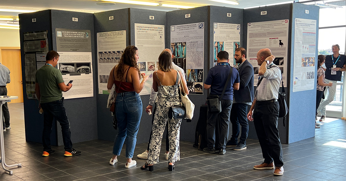
The 22nd Congress of the European Society of Veterinary Orthopaedics and Traumatology (ESVOT) was held from 5 to 7 October at Cruise Terminal 103 in historic Venice, Italy.
ESVOT innovated away from its usual longer multi-stream format to focus on a single area of orthopaedics over two days, which organisers named “In Depth Topic”, with “Complications in Traumatology” being chosen for discussion this year.
It created a compact shared experience for the delegate without the pressures of choice and the worry of missing out on anything.
The choice of complications as a subject is great as, unfortunately, it is something all surgeons experience. So, during a presentation, there can be a delightful tingle of “schadenfreude” when you are not in the hot seat, but this is balanced by one of “freudenfreude” when the colleague who is generously sharing their experience pulls off a success1.
Delegates from Europe and beyond could enjoy various pre-congress labs on 5 October, provided by industry partners who then joined others in the lively commercial exhibition. The main congress programme ran from 6 to 7 October, and was provided by a faculty of very experienced and well-known names, mainly from Europe, providing 14 hours of high-quality CPD.
Avoiding complications firstly involves being aware of those that exist (“the known knowns”, as Donald Rumsfeld put it), so old enemies such as surgical site infection (SSI), septic arthritis, delayed and nonunion were re-explored. Unfortunately, although there are multiple suggested approaches to reduce the incidence of SSIs, proof of their success remains elusive in the literature. Early diagnosis and treatment of a suspect SSI may be aided by sampling for cytology, culture and sensitivity using deep fine-needle aspiration, avoiding any draining tracts.
Septic arthritis is most commonly iatrogenic in origin where it follows recent surgery2. Although prolonged oral antibiotics and local flushing are often effective, one-third of cases may need repeated treatment.
Recent introductions to small animal practice include intra-articular injection of R-gel polymer mixed with amikacin and clindamycin, or intravenous antimicrobial regional limb perfusion to produce above minimum inhibitory concentrations within the target tissues3.
In nonunion cases, autologous cancellous bone graft remains the gold standard as a biological “starting gun” for healing, as it simultaneously provides osteoconductive, osteoinductive and osteogenesis. However, recent research, including ectopic implantation of specific calcium phosphate-based biomaterials, have demonstrated evidence of osteoinduction potential when, previously, they have been seen as just providing an osteoconductive scaffold and a carrier for added osteoinductive factors. It is such a convenient off-the-shelf product, and 3D-printed versions for specific defects with or without additional recombinant human bone morphogenetic protein-2 show promise for veterinary orthopaedic surgeons4.
Presentations looked at fractures that historically have had higher failure rates, such as radial/ulna fractures in toy breed dogs and tibial fractures in cats, which may share biological vulnerabilities such as a reduced vascular network, challengingly limited bone stock (particularly distally) and minimal soft tissue envelopes.
Although the development of more appropriately sized implants and more soft tissue-protecting surgical techniques have reduced the incidence of nonunions in radial/ulna fractures, other complications have filled the vacuum. So, the topic for discussion by Gareth Arthurs (UK)5 and Luca Vezzoni (Italy)6 was loss of bone density after apparently successful healing of the original fracture following dorsal plating, with differing experiences of incidence and approach to management.
One suggestion is destabilisation of the fixation to reduce suspected stress protection by staged removal of the screws starting adjacent to the fracture site and monitoring the response radiographically.
In rare cases, this can lead to the retention of just the plate as a splint stabilised by the ingrowth of bone into the empty screw holes, which creates a “Lego” brick-like interlock. If the original implant is felt to be oversized or possibly too stiff, it can be switched for a better sized and more flexible design. With the screws removed, the stress riser effect from the screw holes disappears as they heal in, reducing the chance of additional fractures while allowing increased loading of the bone simulating its strengthening.
Published work by groups using various external fixator frames, which report high levels of success without encountering the described complication, stimulated discussion as to whether a change of approach is warranted7.
Feline tibial fractures have demonstrated an increased risk of failure, with plate bending in around 13% of cases using medial plates or medial plate-rod combinations. This apparent pattern of acute overload failure has led to recommendations to use orthogonal plating to provide robust fixation.
However, there are certain rules, such as not ending plates at the same level in the diaphyseal rigid cortical portion of the bone, which is poorly able to cope with focal stress concentration compared to the more adaptable metaphysis. Where there is limited bone stock with a very distal fracture, use of a temporary transarticular plate, such as pantarsal arthrodesis plate paired with a cranial plate, can allow the recruitment of extra good bone stock distally, with the arthrodesis plate being maintained for a maximum of six weeks to minimise articular cartilage damage.
Self-knowledge is vital to be aware of how you could react when complications occur. Bruno Peirone (Italy)8 discussed how, so often, things seem to go from bad to worse, and explained this with reference to psychological research on the behaviour of poker players.
Poker players, like surgeons, are skilled, but work with incomplete information in a high-pressure environment that requires constant and rapid decisions to be made. When the game is not going their way, this can lead to the player “tilting”, which means taking increasingly higher risks as they start “chasing” their losses by throwing good money after bad9. Sound familiar? An example could be when unexpected additional haemorrhage during a tibial plateau levelling osteotomy (TPLO) leads to dissection to control it, which damages the cranial tibial artery, causing torrential bleeding with desperate efforts to control this leaving the patient needing a transfusion, a suboptimal osteotomy and maybe a retained swab.
The poker research showed that although the most experienced players felt they “tilted” less, it was found they, in fact, were prone to take more and greater risks, probably based on their expectation of having the opportunity to make any losses back in future games.
Experienced surgeons beware. Surgery can be considered a complex system similar to flying a commercial jet, which is why we have found the transfer of its culture of checklists appropriate. We need to be aware all systems fail, and that complex systems tend to fail in a series of small steps before control is lost.
Therefore, being situational, aware to the warning signs that a cascade is building, will allow the surgeon to stabilise the patient and take time to pause, reset and maybe phone a friend.
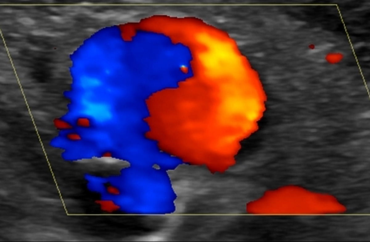
This takes us neatly to a dramatic complication I had never heard of before, which is the development of a large pseudo-aneurysm in two dogs resulting from vascular damage following TPLO surgery, and the placement of an external fixator frame on the proximal tibia. The author, Jean-Guillaume Grand (France)10, discussed that in his two cases, the onset was delayed occurring 25 to 30 days post-surgery.
So, haemorrhage was not suspected until the onset of lethargy, hindlimb lameness and the development of a large pulsatile swelling on the proximal tibia. Colour Doppler ultrasound demonstrated the yin-yang sign, also known as the Pepsi sign, which is the classic appearance of bidirectional blood flow within an aneurysm or pseudo-aneurysm sac7. Treatment involved ligation of the cranial tibial artery and management of the dead space within the swelling, which can be complicated by continuing haemorrhage.
The author suggested the initial damage may have resulted from the projection of a drill bit, saw blade or implant distant from the surgeon’s visible operative field, so not suspected during the operation. So, just as I thought I had heard it all with regard to complications with TPLO surgery, these cases show you can never really relax11.
Newer techniques, such as minimally invasive plate osteosynthesis (MIPO) or arthroscopic-assisted intra-articular fracture reduction, can appear very attractive because of their promises of reduced soft tissue trauma, leading to biological healing and more precise joint reconstruction. However, they also have the risks of new complications, so it is great to hear from experienced pioneers.
Prof Peirone (Italy)12 reported that the most common complications seen in MIPO are mal-alignment of the limb segment, poor fracture reduction and fixation failure. Without sight of the fracture site, the surgeon has to rely on anatomical landmarks at some distance from it, and has to make use of indirect reduction techniques to achieve alignment.
The tibia and radius are more straightforward choices compared to the femur or humerus, and comminuted diaphyseal fractures are more appropriate to the technique compared to so-called simple transverse or oblique fracture patterns.
Mal-alignment may lead to inadequate fixation as implants follow the poorly reconstructed bone, or the difficulties of percutaneous placement through limited portals can lead to the same result. Intraoperative imaging can be extremely useful to avoid the issues described.
Sebastian Knell (Switzerland)13 discussed the pitfalls in arthroscopic-assisted articular fracture repair. Articular fractures require accurate reconstruction of the articular surface and rigid fixation to achieve the best results. However, there is still debate on how much of a step in the articular surface, if any, can be tolerated, and the extent to which degenerative joint disease (DJD) is inevitable following such intra-articular trauma.
Alignment of the joint surface is assessed indirectly via the alignment of extra-articular structures, such as the epicondylar ridges with lateral and medial condylar fractures, using intraoperative imaging with fluoroscopy or on postoperative radiographs. In human articular fracture reconstruction, intra-operative 3D imaging by CT is replacing fluoroscopy, as it produces better radiographic outcomes and more precise implant positioning.

Direct visualisation of fracture reduction is rarely used in veterinary orthopaedics, apart from the occasional use of an approach to the lateral aspect of the humeral condyle and epicondyle, or open examination of the stifle due to the additional trauma produced and limited views available. Arthroscopic assessment of reduction has the promise of a magnified, well-illuminated view of the reduction, and allows assessment of intra-articular structures, but there are potential issues associated with the additional operative time that can be involved, extravasation of fluid and possibilities of additional cartilage damage.
Both speakers warned about the long learning curve associated with these approaches, the need for skilled assistants during surgery, and that surgeons attempting these techniques should be experienced in more traditional trauma surgery so they are able and willing to convert to it, if success is not achieved in a reasonable time frame during the operation.