10 Oct 2016
Simone Anesi discusses the case of a male neutered domestic shorthair cat that presented with a seven-day history of lethargy and tachypnoea, in the latest of Case Notes column.
Simone Anesi
Job Title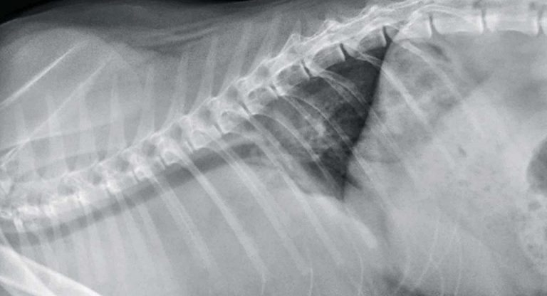
Figure 1. Thoracic radiograph, lateral view.
During a busy morning, Alfie is admitted to your surgery as an emergency. He is a friendly, six-month-old, male neutered domestic shorthair cat that has presented with a seven-day history of lethargy and tachypnoea.

He is quiet, but alert and responsive in the consult room. Your clinical examination reveals marked enlargement of his prescapular and popliteal lymph nodes, and his respiratory rate is 60 breaths per minute with moderate inspiratory effort. Orthogonal conscious thoracic radiographs are acquired (Figures 1 and 2).
What do these radiographs show?
Mild rotation exists of the spine in both projections and the forelimb triceps musculature is partially superimposed over the cranial thorax on the lateral view. Wet coat artifact is also evident on dorsoventral projection. Both radiographs reveal a marked increase in soft tissue opacity predominantly within the cranial aspect of the thorax resulting in border effacement of the cranial cardiac silhouette.

Notably, on the dorsoventral projection, evidence exists of marked widening of the cranial mediastinum. On both views, only the caudal margin of the heart is visible and indicates the heart is displaced caudally, and to the right of midline within the thorax.
The size of the heart cannot be accurately assessed from these radiographs. There is evidence of elevation of the entire intrathoracic trachea and the tracheal carina has been displaced caudally from its normal position around the fifth intercostal space, to the seventh intercostal space.
On the dorsoventral projection there is also the impression of increased soft tissue opacity cranial to each scapulohumeral joint, most likely due to marked enlargement of the prescapular lymph nodes. Skeletal structures are within normal limits.
In summary, the key radiographic findings are marked widening of the cranial mediastinum and caudal and dorsal displacement of the trachea. Although tracheal elevation may be caused by a large cranial mediastinal mass, large volume of pleural fluid or heart base mass, only a cranial mediastinal mass would explain caudal displacement of the tracheal carina.
Several differential diagnoses exist for a cranial mediastinal mass in cats, including mediastinal neoplasia (such as lymphoma or thymoma), a mediastinal cyst or, very rarely, ectopic thyroid tissue, granuloma, haematoma and abscess. The presence of concurrent peripheral lymphadenopathy makes lymphoma the most likely diagnosis.

Thoracic ultrasound was performed and confirmed the presence of a large soft tissue mass within the cranial mediastinum. The mass had a complex loculated appearance and contained several pockets of hyperechoic fluid (Figure 3).
Fine needle aspiration of the prescapular lymph nodes was performed and submitted to an external laboratory. The sample was characterised by a predominantly lymphoid population consistent with high-grade lymphoma.
Following his diagnosis, Alfie’s owners were keen to pursue treatment. He was hospitalised for several days, during which time chemotherapy involving vincristine, cyclophosphamide and prednisolone was administered. His tachypnoea gradually improved and eventually resolved completely within a week. His enlarged peripheral lymph nodes also returned to normal size.
Alfie’s demeanour improved and, six months after presentation, his owners reported he was tolerating his chemotherapy well and enjoying an excellent quality of life.