19 Oct 2022
Francesco Cian DVM, DipECVCP, FRCPath, MRCVS presents the case of a five-month-old boxer with a skin lesion in the latest of his Vet Times Diagnostic Dilemmas.
Francesco Cian
Job Title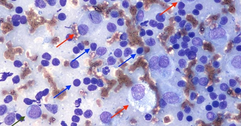
Figure 1. Fine needle aspirate from a skin mass in a dog (Wright-Giemsa, 50×).
A five-month-old boxer presented for recent onset of a small, raised, dome-shaped skin lesion on the head. The veterinarian performed a fine needle aspirate of the lesion and submitted the slides to an external laboratory for cytological examination.
The submitted smears (Figure 1) are highly cellular with adequate preservation. The background is lightly basophilic with red blood cells, a few bare nuclei and lymphoglandular bodies (cytoplasmic fragments of lymphoid cells).
A main population of small lymphocytes is present (blue arrows), characterised by small, hyperchromic round nuclei and basophilic cytoplasm rim. They are admixed with a few intermediate-size lymphoid cells (green arrows) and a few histiocytic cells (red arrows). These cells have moderate to abundant amounts of lightly basophilic cytoplasm, with defined borders and occasionally vacuolated.
Nuclei are round – often multiple – with coarse, granular chromatin and visible nucleoli. Anisocytosis and anisokaryosis are moderate.
A diagnosis of regressing cutaneous histiocytoma was determined.
Cutaneous histiocytoma is a common benign neoplasm originating from Langerhans cells – histiocytic cells of the epidermis. It often appears as a solitary, dome or button-shaped dermal lesion, variable in size, sometimes bright red in colour, rapidly growing and prone to ulceration.
Multiple lesions are uncommon and reported mainly in Shar Pei dogs. Histiocytoma is typically found on the head (including the ear pinna), neck or limbs of young dogs, and certain breeds (for example, the boxer and dachshund) seem to be predisposed. However, histiocytoma may occur in dogs of all breeds and ages. It has not been reported in cats.
Cytologically, cutaneous histiocytoma is characterised by a proliferation of histiocytic cells, which may show features of atypia (for example, including bi-multinucleation, anisocytosis/anisokaryosis and mitotic figures), despite its benign behaviour. In mature lesions, it is not uncommon to observe an infiltrate of small lymphocytes representing the T-cell immune response, often leading to a spontaneous regression of the lesion within a few months.
The heavy lymphocytic infiltrate that occurs in late-stage regression of histiocytomas can be mistaken on cytology for small cell lymphoma – especially in the absence of histiocytic cells. Flow cytometry or molecular clonality testing are non-invasive diagnostic procedures that are expected to resolve this issue.
However, clonal expansions of CD8+ lymphocytes have been documented in association with the cytotoxic T-cell response to histiocytoma and these findings are not different from what is observed in T-cell lymphoma. Therefore, in the presence of dubious lesions that do not resolve spontaneously, biopsy and histopathological examination should be preferred for a definitive diagnosis.