8 Dec 2020
In a Case Notes piece from Vet Times, Ivan Filipovic PgC(SAS), MRCVS looks at the case of acute onset non‑weightbearing lameness of the right thoracic limb.
Ivan Filipovic
Job Title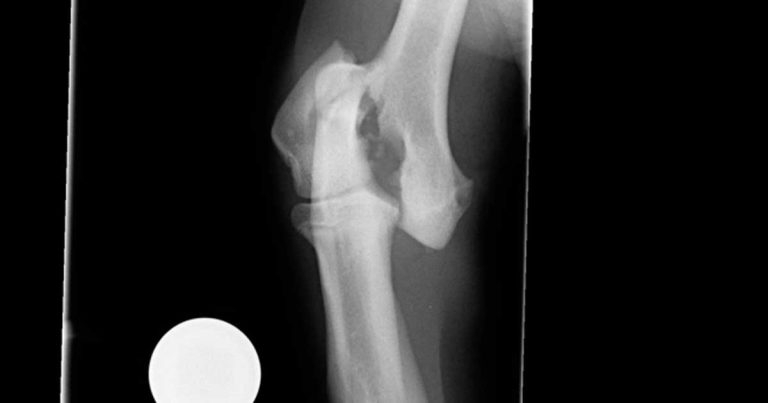
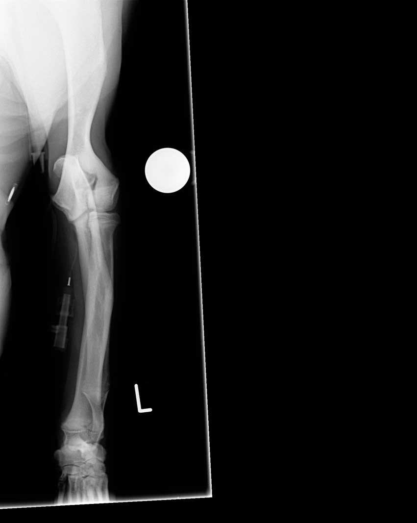
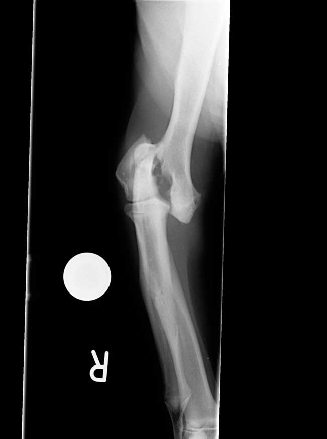
Figure 1. Preoperative craniocaudal radiograph of the right elbow.
During a busy morning surgery you are presented with Hugo, a seven‑month‑old, male springer spaniel, with an acute onset non‑weightbearing lameness of the right thoracic limb that occurred while chasing a ball.
Previous intermittent left thoracic limb lameness had also been noted; this had been treated conservatively with NSAIDs.
Orthopaedic examination revealed swelling, crepitus and pain associated with the right elbow, and mild discomfort on firm extension of the left elbow.
As a first step of investigation and treatment, you provide analgesia and perform radiography of the right elbow (Figure 1) under general anaesthesia.
A fracture of the lateral aspect of the humeral condyle is apparent. What would you do next?
The radiograph confirmed a fracture of the lateral aspect of the humeral condyle on the right side. However, the patient’s history is also suggestive of chronic lameness and discomfort associated with the contralateral limb. Further investigations are, therefore, indicated prior to proceeding with surgery.
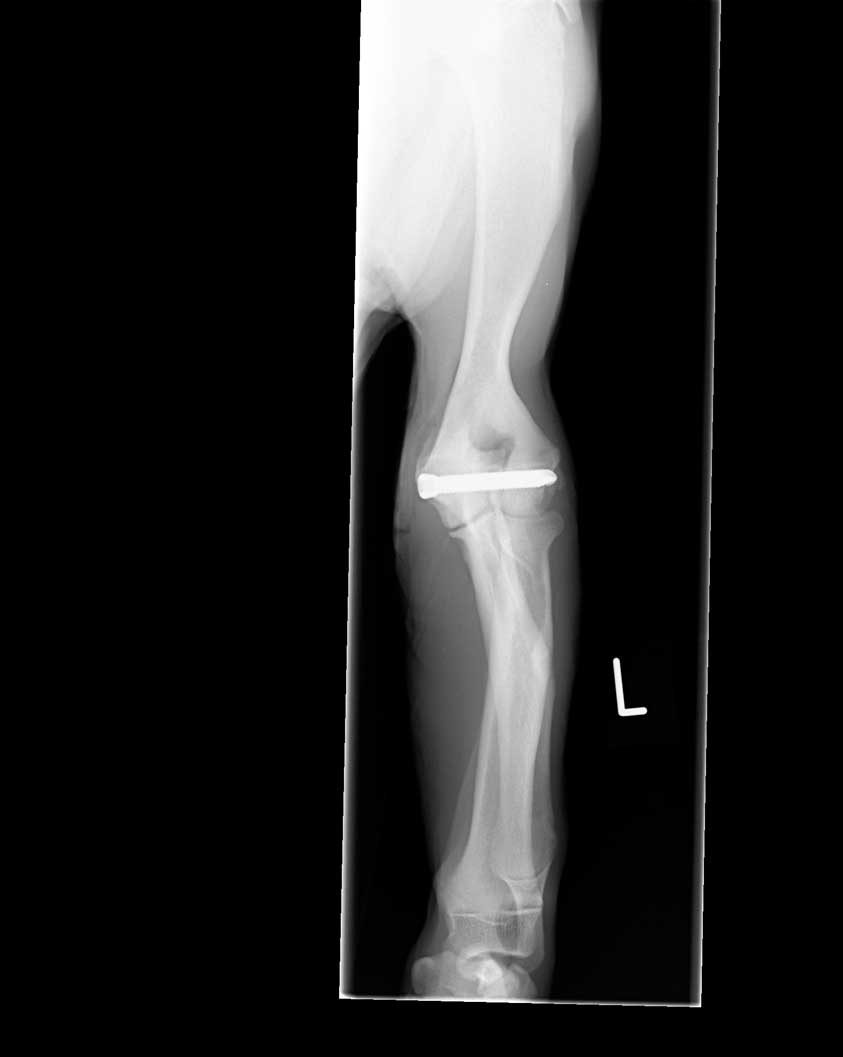
Additional radiographs were taken of the left elbow. On a craniocaudal view a humeral condylar fissure was apparent (Figure 2).
Fracture of the lateral part of the humeral condyle is most often associated with low‑energy trauma, such as jumping, and mainly affect patients younger than one year of age, with a peak at four months of age. If this fracture is seen in older dogs, particularly after minimal trauma, an underlying humeral condylar fissure should be considered.
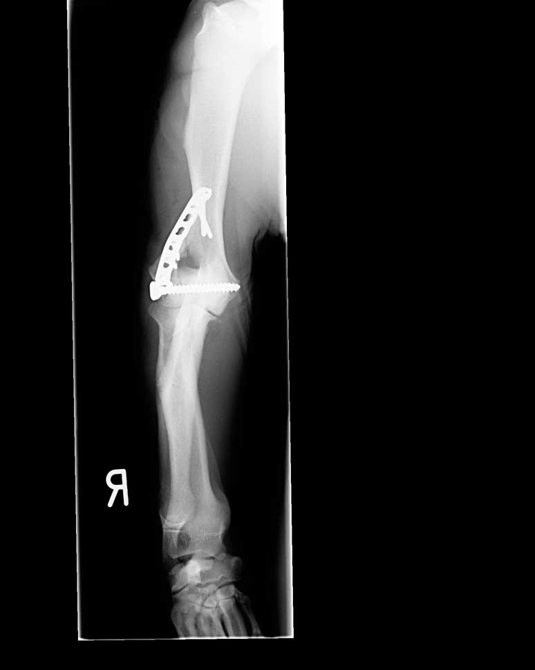
This patient’s fracture was treated by open reduction and stabilisation using a 4.5mm transcondylar lag screw and 2.4mm locking compression plate, applied with a combination of locking and non-locking screws (Figure 3).
Since a humeral condylar fissure was confirmed on the left side, a decision was made to place a 5mm transcondylar screw in a medial to lateral direction (Figure 4). The purpose of this screw was to reduce discomfort associated with the underlying fissure and reduce the likelihood of the dog suffering a condylar fracture.
The dog had an uneventful recovery and was sound at examination six weeks post‑surgery.
A retrospective study of patients with humeral condylar fissures treated with lateral to medial transcondylar screws found an overall complication rate of 59.5%, with seroma and surgical site infection being recognised most commonly, and implant failure seen less frequently (Hattersley et al, 2011).
Conversely, a study by Clarke et al (2012) found a much lower complication rate when screws were placed in a medial to lateral direction.
This case illustrates the importance of obtaining radiographs of the contralateral limb as well as the fractured limb, particularly in dogs with a history of prodromal lameness.
This article was reviewed by Toby Gemmill BVSc, MVM, DSAS(Orth), DECVS, FRCVS.