4 Sept 2017
Mahy Rodriguez Blanco discusses the consult of Enzo, a six-year-old male, neutered, cross-breed rescue dog, with a left forelimb lameness.
Mahy Rodriguez Blanco
Job Title
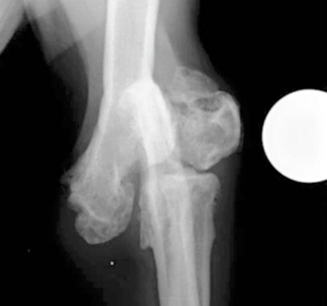
Orthogonal radiograph of the left elbow, showing a chronic lateral humeral condylar fracture malunion.
Your first consult on a busy Monday morning is Enzo, a six-year-old male, neutered, cross-breed dog, rescued two weeks earlier by his new owners.
The owners report Enzo has had a left forelimb lameness; however, he has been happy and able to go on his normal walks, run freely and play with other dogs.
On examination, Enzo is bright and alert, with a normal rectal temperature. Orthopaedic examination confirmed a 4/10 left forelimb lameness with moderate associated muscle atrophy. The left elbow is thickened with a decreased range of motion, and mild discomfort is apparent on manipulation of the joint. In addition, a suspicion of subtle discomfort is present on firm extension of the right elbow.
The patient was sedated and orthogonal radiographic projections of the left elbow obtained. These demonstrate a chronic lateral humeral condylar fracture malunion (Figure 1).
What would your next step be?
Malunions are the result of a fracture where, due to an inappropriate treatment, bone healing occurs, but with a degree of deformity and malalignment. Careful clinical assessment is required since malunions can be either functional – in which case no treatment is required – or non-functional, in which case surgery should be considered. Surgery to improve alignment can be challenging; therefore, the relative benefits and risks of conservative versus surgical treatment should be considered. In extreme cases, limb amputation may be an appropriate option.
In Enzo’s case, conservative treatment of the left forelimb was considered the best option, as surgical treatment was deemed unlikely to have improved the limb function and could have created significant morbidity.
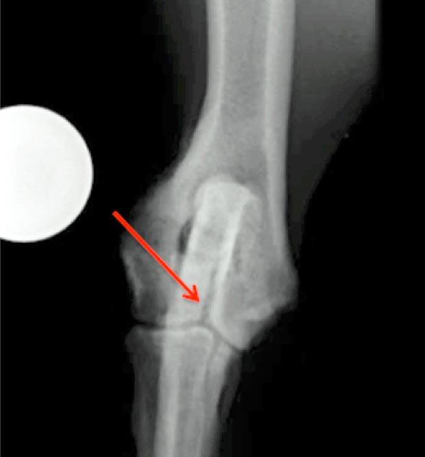
Since Enzo had a humeral condylar fracture, assessment of the contralateral elbow was indicated, as the presence of a humeral condylar fissure can predispose to complete condylar fractures, particularly in the spaniel breed. A craniocaudal radiograph of the right elbow (Figure 2) confirmed the presence of a fissure.
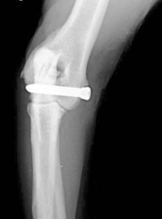
Since suspicion of pain was present on manipulation of the right elbow, and because there was an increased risk of progression of the fissure to a complete fracture in the future, placement of a transcondylar screw was undertaken (Figure 3).
A 5mm screw was used, with a core diameter of 4.4mm, reducing the risk of long-term implant failure compared to smaller screws. The screw was placed in a medial to lateral direction, which has been shown to decrease the risk of postoperative complications, such as seroma formation and infection.
Enzo recovered uneventfully following surgery and, six months following surgery, the owners reported excellent right forelimb function. As expected, left forelimb lameness had persisted, although this had reportedly improved with conservative treatment.