17 Jul 2017
The two images are from an aspirate from a large shoulder mass in an adult dog. The author asks: "What is your diagnosis?"
Francesco Cian
Job Title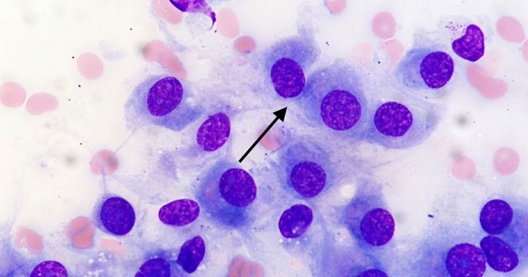
The cytology pictures from an aspirate from a large shoulder mass in an adult dog.
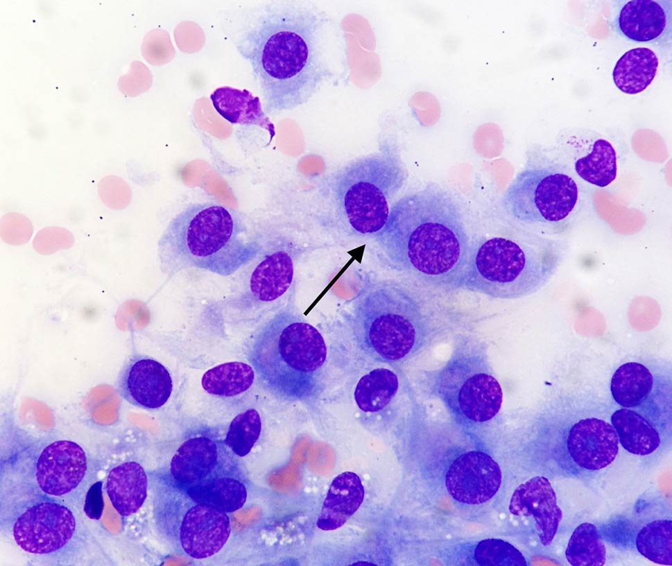
The two images on the right are from an aspirate from a large shoulder mass in an adult dog (Wright-Giemsa, 20× to 100×). What is your diagnosis?
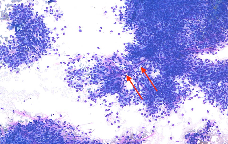
The aspirate is slightly haemodiluted and harvested high numbers of well-preserved nucleated cells on a clear background. These cells appear slightly elongated and tend to form bundles adherent to the surface of capillaries (elongated pinkish structures; red arrows).
Cells (black arrow) have moderate amounts of basophilic cytoplasm, occasionally elongated, with poorly defined borders and rarely contain small numbers of intracytoplasmic clear vacuoles and scattered light pink granules.
Nuclei are round to oval shaped and paracentrally located, with granular chromatin and poorly distinct, round nucleoli. Anisocytosis (cell size variation) and anisokaryosis (nuclear size variation) are mild.
Rare binucleated and scattered multinucleated cells – the latter, known as crown cells, and not seen in these images – are also noted throughout the smears, together with rare neutrophils, likely blood derived.
These findings are consistent with a diagnosis of perivascular wall tumour.
Perivascular wall tumours (previously called haemangiopericytoma) are relatively common skin neoplasms affecting the canine species – in particular, middle-aged or older subjects. Tumours with a similar morphology occur only rarely in cats.
Studies have shown this is not one neoplasm, but represents a spectrum of tumours arising from various cells of the perivascular wall and the adventitia (for example, pericytes and myopericytes), including:
They often appear as solitary lesions, with predilection for the limbs and joints. They have variable gross appearance and sometimes may be confused for lipoma, given their rubbery to fatty appearance.
Treatment involves wide surgical excision (where possible) and is often curative. Local recurrence is common, but metastatic risk is relatively low.
Histopathological examination of these lesions is also recommended and helpful to further confirm the diagnosis. However, of all the spindle cell tumours, this one exfoliates the greatest number of neoplastic cells, making a cytological diagnosis relatively easy.
Peripheral nerve sheath tumours and soft tissue sarcomas of different origin should also be considered as possible, less likely differentials,since they may share similar cytological features.