25 Feb 2019
Mark Lowrie discusses diagnosis of this chronic condition, the wide range of drugs and alternative therapies, and why it is vital to efficiently deal with owners’ expectations.
Mark Lowrie
Job Title
Figure 1. Indications to perform MRI include abnormal behaviour between seizures, an abnormal neurological examination and failure to respond appropriately to antiepileptic medication.
Idiopathic epilepsy requires careful management that begins with precise diagnosis, accurate administration of medication and – probably most importantly – management of client expectations.
Given up to a third of dogs with epilepsy can become refractory (fail to respond appropriately to adequate dosages of conventional medication), inadequate control – despite a veterinarian’s best efforts – is common. Therefore, client compliance can become increasingly important.
Consultations often begin with an owner declaring “my pet has had a seizure”. This assumption should be challenged.
Paroxysmal episodes are difficult to characterise because, by definition, they are episodic with long periods of normality. The paroxysmal nature of these events, combined with the possible presence of prodromal or postictal changes, is usually suggestive of a neurological condition and makes many owners assume a seizure.
However, with the advent of the smartphone, it is now possible to gain an insight into what the owner has observed. This has revolutionised the way vets manage dogs and cats with paroxysmal episodes and, although a description may appear diagnostic of an epileptic seizure, the reality is often different.
Signs that are useful in distinguishing epileptic seizures from other paroxysmal disorders include the presence of autonomic signs – for example, urination, defecation and salivation – loss of awareness, and the presence of abnormal behaviour before or after each event (prodromal or postictal phase, respectively).
Other signs that may help, but are not necessarily diagnostic of an epileptic seizure, include the presence of increased tone, stereotypical episodes (each event looks the same), abrupt onset and termination (except reflex seizures) and a short duration of the episode itself (usually shorter than a few minutes).
The presence of decreased tone during a paroxysmal episode is strongly suggestive of a non-epileptic seizure disorder, while a long duration – for example, 30 to 60 minutes, or longer – would also suggest an epileptic seizure is less likely, particularly when accompanied with no postictal signs.
If clinical signs of forebrain disease are present, it is imperative a neurological examination is performed, as abnormal findings lend support for metabolic or symptomatic causes for seizures.
However, in the absence of these signs, structural forebrain disease cannot be excluded. Certain aspects of the neurological examination are particularly important in assessing forebrain function:
Extracranial causes are also termed “reactive” seizures. These include metabolic diseases (such as hypoglycaemia, hypocalcaemia and hepatic encephalopathy) and toxicities (for example, metaldehyde, permethrin, organophosphates and mycotoxins).
Clinical pathology testing should include a complete blood count, fasted serum biochemistry – consisting of glucose, sodium and calcium concentrations, and renal and hepatic dysfunction (notably urea, creatinine, cholesterol and albumin) – and urinalysis. Abnormal findings may further support a metabolic or toxic cause of seizures.
Based on clinicopathological abnormalities, additional diagnostic testing is indicated when specific organ pathology is suspected. Liver function tests – such as preprandial and postprandial bile acid concentrations, and blood ammonia concentration – can provide evidence of hepatic dysfunction.
If extracranial disease is ruled out then an intracranial problem is suspected. Additional testing can further determine the type and extent of intracranial pathology.
Cross-sectional imaging, such as CT or MRI, is the gold standard at identifying intracranial lesions – with MRI (Figure 1) preferred due to its better sensitivity for identifying soft tissue structures.
The indications to perform MRI are detailed in Panel 1.
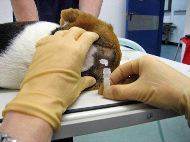
CSF analysis is particularly useful in identifying the presence of inflammatory CNS disease (Figure 2).
However, results of this evaluation are rarely pathognomonic for a specific aetiology when performed in isolation, and are best interpreted in all patients alongside cross-sectional imaging findings.
Doing this can give a clearer indication of whether an inflammatory or non-inflammatory process is present, and – in light of signalment, history and other findings (for example, cross-sectional imaging and serology) – leads to refinement of the diagnosis.
Serology and immunological testing may indirectly lend further support to infectious causes.
Overall, viral causes are difficult to definitively diagnose and the results of serological testing are difficult to interpret because of the presence of circulating antibodies from maternal immunity, vaccination or environmental exposure.
Serology is, therefore, best performed in combination with intracranial imaging – rather than as a standalone test – to avoid misinterpretation of the results.
Lifelong medication is often necessary in dogs with idiopathic epilepsy.
The use of the word treatment is often confusing to owners, as it implies a resolution of seizures. In reality, management of seizures is the only realistic goal and the response to antiepileptic drugs is not always satisfactory. Furthermore, antiepileptic drugs can cause transient, dose-related or idiosyncratic side effects that may be more undesirable than the seizures.
The most appropriate choice of antiepileptic drug for one animal may not be the best choice for another. When recommending a new or alternative treatment for epilepsy, it is important to provide owners with unbiased information of the expected effectiveness of the particular treatment, as well as some of the practicalities – for example, the cost and frequency of dosing.
Some of this information is simple and straightforward, but the major question of whether the drug is effective can be very difficult – if not impossible – to answer, as controlled studies are not available for many of the newer medications.
The majority of data on drug efficacy presented in this article have been obtained from studies in which the antiepileptic drug was administered in an open label fashion. Generally, dogs with “poorly controlled” seizures (often described as refractory epilepsy) were given a novel antiepileptic drug, and seizure frequency during administration of the drug was compared with the seizure frequency at baseline.
A reduction in seizure frequency has been associated with placebo administration in dogs with epilepsy (Muñana et al, 2010). Almost 30% of the dogs in this study were classified as responders to placebo (response defined as 50% or greater reduction in seizure frequency).
Two reasons that may have explained this phenomenon were given. Firstly, epilepsy is a waxing and waning disorder; therefore, normal, unpredictable fluctuations in seizure frequency occur over time. Owing to the cyclical nature of the disease, an improvement in seizure frequency is likely regardless of the management regimen adopted, and this improvement may be mistakenly attributed to a recently instituted change in therapy.
A second factor that may play a role in this “placebo effect” is that owners enrolling their dog in a study of a novel antiepileptic drug may perceive an improvement simply through the additional attention provided by the investigators running the study.
A study evaluating the use of the anticonvulsant levetiracetam demonstrated a significant reduction in seizure frequency, in all patients during the study period, that was independent of the treatment type (levetiracetam or placebo), as well as an improvement in owner-perceived quality of life, which supports this hypothesis (Muñana et al, 2010).
Open-label studies cannot account for this bias; therefore, the efficacy reported in the majority of veterinary studies of novel antiepileptic drugs may be exaggerated. The bottom line is the efficacy of many novel antiepileptic drugs remains uncertain, and clients should be made aware of this.
Phenobarbital is a barbiturate traditionally used as a monotherapy for seizures in dogs and cats.
Phenobarbital exerts its antiepileptic effect by enhancing the conductivity of the gamma-aminobutyric acid (GABA)-chloride channel complexes, thereby reducing both glutamate-mediated excitation and calcium ion flow into neurons.
The initial dose is 2mg/kg to 3mg/kg twice daily, and serum concentrations reach a steady state within 10 to 14 days. Therefore, it is advisable to check serum concentrations 2 weeks after starting treatment and to collect samples at consistent times after administration.
To maintain therapeutic serum concentrations, subsequent dose increases may be required. This is because of the induction of hepatic enzymes leading to a reduction in the elimination half-life of the drug.
Sedation and ataxia are common adverse effects associated with this medication, although they are usually transient and only persist in some cases. Polyphagia, polydipsia and polyuria are also frequently encountered, and are usually dose-related.
Other side effects associated with phenobarbital are classified as idiosyncratic, as they seem to be unrelated to dose and prolonged administration. These include hyperexcitability, acute hepatotoxicity, blood dyscrasias – for example, thrombocytopenia, anaemia or leukopenia – and superficial necrolytic dermatitis.
Hepatotoxicity has been suggested to be dose-related – in one study, it was found dogs with hepatotoxicity had phenobarbital concentrations exceeding 40µg/ml (Müller et al, 2000).
It is also important to understand the normal laboratory changes induced by phenobarbital. The most common changes observed are asymptomatic, mild-to-moderate elevations in serum levels of alkaline phosphatase and alanine transaminase. However, these increases cannot be used solely as a signal of underlying liver disease; therefore, a bile acid stimulation test is recommended as routine monitoring for dogs on phenobarbital.
Imepitoin is a novel antiepileptic drug licensed for monotherapy in first-line treatment of idiopathic epilepsy in dogs, although available information on its relative efficacy and long-term side effects in large numbers of dogs is limited.
Imepitoin has a half-life of approximately 1.5 hours (although this is dose-dependent, and can vary at higher and lower doses), with steady state being achieved rapidly within 3 days. It is metabolised via oxidative metabolism in the liver. The cytochrome P450 system is not involved in imepitoin metabolism; therefore, it does not induce liver enzymes with long-term administration of the drug in the way phenobarbital does.
Reported transient side effects during the first weeks of treatment include excitability, hyperactivity, sedation, ataxia, polyphagia, mild generalised tremors, gastrointestinal disorders and an increased incidence of aggression. Imepitoin does not require serum concentration monitoring and withdrawal seizures have not been described, although careful withdrawal of the medication is recommended if required.
Imepitoin is contraindicated in cluster seizures and status epilepticus.
Potassium bromide is effective as a monotherapy, but is most commonly used as an add-on therapy.
According to the results of one study, approximately one in four dogs with epilepsy resistant to phenobarbital treatment achieve a seizure frequency reduction of 50% or more when potassium bromide is added to the management regimen.
Bromide competes with chloride at GABA receptors, hyperpolarising the neuronal membrane and increasing the seizure threshold. Bromide also competes with chloride for renal elimination; therefore, sodium chloride intake must not be altered in dogs receiving this medication. Failure to achieve this will result in increased elimination of bromide, or bromide toxicosis. Renal insufficiency also decreases bromide elimination; therefore, potassium bromide should be used cautiously in patients with this condition.
A disadvantage of using potassium bromide is its long elimination half-life of approximately 25 days. A steady-state concentration is achieved only after three to four months of initiating therapy – this is important to remember when measuring the serum concentration, as blood sampling before this time may indicate a subtherapeutic serum concentration.
The reported side effects of potassium bromide include vomiting, weight gain, polyphagia, pancreatitis, polyuria and polydipsia.
Levetiracetam is one of the more recently approved antiepileptic drugs for humans and is probably one of the more commonly used medications in the management of refractory canine epilepsy.
Although generally recommended as an add-on antiepileptic drug, it has been used successfully in humans as monotherapy. Its mechanism of action has not been fully elucidated and some controversy exists as to whether it may reduce the risk of seizure-induced brain damage.
Levetiracetam has minimal hepatic metabolism in dogs, with more than 80% of the drug excreted in the urine, and the remainder being hydrolysed in the serum and other organs.
The half-life in dogs is 3 to 4 hours, which necessitates frequent administration and means a steady-state concentration is reached within 48 hours. The recommended oral dose is 20mg/kg every 8 hours.
No clear understanding exists of the relationship between serum drug concentration and efficacy; therefore, serum monitoring is not performed for this medication.
The proposed benefits of a hypoallergenic diet are derived from the avoidance of toxins or allergens that may “trigger” seizures (Collins, 1994).
Human studies have reported an unusually high incidence of allergic disease among patients with epilepsy (more than 50%) and a pilot study has investigated a similar phenomenon in dogs (Luján et al, 2004). This study included eight dogs with treatment-refractory epilepsy – of these, seven were found to have gastrointestinal or skin allergies in conjunction with their seizures. The introduction of an exclusion diet reduced seizures to an “acceptable level” in seven of eight dogs. Behavioural abnormalities associated with seizures were completely eliminated in all cases.
Nothing further has been reported since this trial, but anecdotal reports support the idea of incorporating a hypoallergenic food trial into the regimen of dogs with refractory epilepsy.
The rationale behind the use of a ketogenic diet has been extrapolated from studies in humans that indicate inducing ketosis can decrease seizure frequency in children.
However, differences in metabolism between humans and dogs mean the levels of ketosis easily induced in humans cannot be induced by dietary manipulation in dogs. Furthermore, the results of a ketogenic food trial in dogs with idiopathic epilepsy failed to demonstrate a difference in seizure frequency between dogs that received a high-fat, low-carbohydrate diet and those that received a control diet, although the trial was limited in that the number of dogs enrolled was small (Patterson et al, 2005). Therefore, ketogenic diets have not found a role in the management of canine epilepsy.
One study did investigate the use of medium-chain triglyceride diet for canine epilepsy. The six-month prospective, randomised, double-blind, placebo-controlled, crossover dietary trial was designed to compare a ketogenic medium-chain triglyceride diet with a standardised placebo diet in dogs with refractory idiopathic epilepsy. Twenty-one dogs were fed one of the diets for three months then switched to the other diet for a further three months, with seizure frequency recorded.
Seizure frequency was significantly lower when dogs were fed the ketogenic diet (2.31 seizures per month) compared with the placebo diet (2.67 seizures per month). Of the 21 participating dogs, 3 dogs became seizure-free and 7 additional dogs had more than a 50% reduction in seizures (Law et al, 2015).
In humans with epilepsy, fatty acid supplementation has been found to alleviate seizures and some of the symptoms associated with epilepsy – for example, behavioural change.
In one study, the authors enrolled 15 dogs with idiopathic epilepsy that had a poor response to conventional medication (Matthews et al, 2012). A unique feature of this study was it was one of the first studies in canine epilepsy to include a double-blind, placebo-controlled, crossover methodology.
Each dog had a 12-week trial phase in which they received an essential fatty acid supplement or placebo; after this, the treatments were swapped and continued for a further 12 weeks. The owners were unaware of which trial phase the dog was in.
Although no difference in seizure frequency was observed between the fatty acid supplement recipients and placebo recipients, one dog had a dramatic reduction in seizures when receiving the fatty acid supplement, while another had a marked reduction in aggressive behaviour. Unfortunately, the number of cases and the short trial period was such that no significant conclusion could be drawn.
The wide variety of antiepileptic drugs and alternative therapies available for the treatment of canine idiopathic epilepsy simply reveals the lack of understanding of the condition.
Every patient with idiopathic epilepsy should be treated on an individual basis, as the correct choice for one dog may be entirely inappropriate for another. The recommended way to choose the most appropriate antiepileptic drugs is to become familiar with the reported efficacy, side effects and metabolism of each medication, to give owners an informed choice on the advantages and disadvantages of each.
It is expected further placebo-controlled studies will soon be available for many of these medications.